Search results
From Embryology
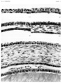
File:Streeter1957 plate01.jpg A segment of the cornea near the mid-line is shown in an embryo of each horizon. The mesoclermal component is a thin layer one or two cells Fig. 14. [[:Category:Carnegie Embryo 5609|'''No. 5609''']], xix, I5-I-I x 800.(1,500 × 2,009 (486 KB)) - 17:37, 6 November 2016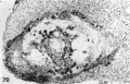
File:Hertig1956 fig70.jpg ==Fig. 70. Abnormal Implanted Ovum== ...to lack of or early death of the embryo. [[:Category:Carnegie Embryo 7771|Carnegie 7771]], Section 34-1. X 100.(1,124 × 723 (157 KB)) - 13:32, 25 February 2017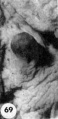
File:Hertig1956 fig69.jpg ...ugh there were many other polyps present. [[:Category:Carnegie Embryo 7771|Carnegie 7771]], Sequence 1. X 1-1.(360 × 738 (31 KB)) - 13:29, 25 February 2017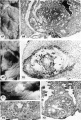
File:Hertig1956 plate13.jpg ...nd a small chorionic cavity but no embryo and a third with trophoblast and embryo but no chorionic cavity. It is, perhaps, conceivable that abnormal morulae ...ial wrinkling of its endometrial surface. [[:Category:Carnegie Embryo 8329|Carnegie 8329]], Sequence 2. X 22.(1,280 × 1,896 (487 KB)) - 15:35, 25 February 2017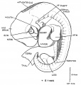
File:Keith1921 fig044.jpg ==Fig. 44. Outline of a Human Embryo 10.4 mm long and in the 6th week of development== ..., Ibid. p. 492. Dr. H. L. Bamiville gives a full description of an 8-5 mm. Embryo, Journ. Anat. 1915, vol. 49, p. 1. Dr. F. W. Thyng gives excellent figures(1,027 × 1,079 (144 KB)) - 15:44, 24 May 2017


