Search results
From Embryology
File:Sensenig1951 plate02.jpg Fig. 7, 8. [[Carnegie Collection|Carnegie Embryo]] no.{{CE8172}} age group xviii Fig. 9, 10. [[Carnegie Collection|Carnegie Embryo]] no.{{CE7274}} age group xx(2,078 × 2,619 (1.52 MB)) - 19:16, 27 March 2018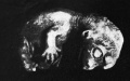
File:Mall1908a plate03fig08.jpg ==Plate III Fig. 8. Normal human embryo No. {{CE256}} 16 mm long== | Embryo {{CE256}} was later classified as [[Carnegie stage 20]].(1,106 × 694 (43 KB)) - 09:53, 1 August 2018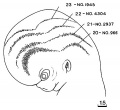
File:ORahilly1987 fig20-2.jpg ==Fig. 20-2. Head Superficial Vascular Plexus - Stages 20-23== ...showing the edge of the superficial vascular plexus in the head at stages 20-23.(600 × 549 (46 KB)) - 13:28, 22 May 2017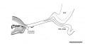
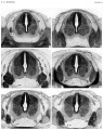
File:Streeter1957 fig04-20.jpg ==Fig. 4. Drawing of sections through the eye and optic nerve in Stage 20== Several sections in each embryo were combined to show the form and dimensions of the optic nerve. x50.(1,538 × 800 (78 KB)) - 23:00, 17 April 2018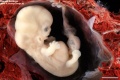
File:Stage20 bf11.jpg ==Human Embryo Carnegie Stage 20== Appears to be an [[Carnegie stage 20|early Stage 20 embryo]].(1,200 × 800 (172 KB)) - 09:54, 18 March 2014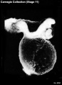
File:Stage11 bf16.jpg ==Human Embryo Carnegie Stage 11== Carnegie Collection Embryo No.6050(420 × 577 (27 KB)) - 13:21, 22 April 2012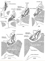
File:Streeter1957 fig08.jpg File:Streeter1957 fig08-19.jpg|[[Carnegie stage 19|stage 19]] (Embryo {{CE1390}}) File:Streeter1957 fig08-20.jpg|[[Carnegie stage 20|stage 20]] (Embryo {{CE6202}})(1,280 × 1,712 (507 KB)) - 10:16, 23 May 2017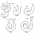
File:Streeter1957 fig07.jpg ...cimens used are members of the respective age groups with the exception of embryo no. 86, which is slightly more advanced in bone-marrow formation and is not ...er1957 fig7-19.jpg|[[Carnegie stage 19]] ([[:Category:Carnegie Embryo 5609|Embryo 5609]])(1,280 × 1,201 (117 KB)) - 20:43, 15 March 2017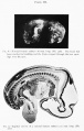
File:Mall1908a plate03.jpg | Fig. 8. Normal human embryo 16 mm. long (No. {{CE256}}). The head had been crushed in handling and the | Fig. 9, Sagittal section of a normal human embryo 7.5 mm. long (No. {{CE221}}).(1,743 × 2,738 (289 KB)) - 10:04, 1 August 2018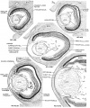
File:Streeter1957 fig06.jpg ...er1957 fig06-19.jpg|[[Carnegie stage 19]] ([[:Category:Carnegie Embryo 409|Embryo 409]]) ...er1957 fig06-20.jpg|[[Carnegie stage 20]] ([[:Category:Carnegie Embryo 462|Embryo 462]])(1,280 × 1,541 (622 KB)) - 13:41, 22 May 2017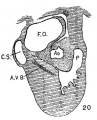
File:Mall1912-fig20.jpg ==Fig. 20. Sagittal section Embryo 15.5 mm long== Carnegie Embryo No. {{CE390}} x 18.(400 × 494 (56 KB)) - 16:21, 7 November 2017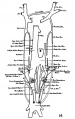
File:McClure1925 fig14.jpg ==Fig. 14 Diagram of the venous system of a 22 mm human embryo== {{Online Editor}} - This embryo could be either [[Carnegie stage 20]] or [[Carnegie stage 21]], both stages occur in [[Week 8]].(1,000 × 1,586 (211 KB)) - 10:04, 19 January 2017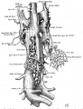
File:McClure1925 fig15.jpg ==Fig. 15 Reconstruction of the venous system of a 22 mm human embryo== {{Online Editor}} - This embryo could be either [[Carnegie stage 20]] or [[Carnegie stage 21]], both stages occur in [[Week 8]].(1,000 × 1,301 (309 KB)) - 10:07, 19 January 2017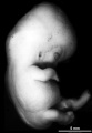
File:Bl130758-01.jpg ==Human Embryo (19.2 mm)== [[Carnegie stage 20]] [[Week 8]](753 × 1,091 (59 KB)) - 13:58, 1 September 2015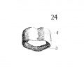
File:Lisser1911 fig24.jpg ...aphic reconstruction of cricoid cartilage posterior veiw in human Carnegie Embryo 22== [[:Category:Carnegie Embryo 22|'''Embryo no. 22''']] (20 mm.) 3, anterior arch; J!^, posterior arch.(826 × 705 (36 KB)) - 21:54, 15 June 2016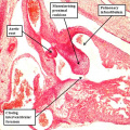
File:Anderson2016-fig48.jpg ==Fig. 48. Human Heart (Carnegie stage 20)== The section is taken from a human embryo at [[Carnegie stage 20]], in the eighth week of development, just prior to closure of the embryoni(800 × 800 (203 KB)) - 21:53, 16 February 2017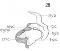
File:Lisser1911 fig28.jpg ==Fig. 28. Graphic reconstruction of laryngeal cartilages in human Embryo no. 22== [[:Category:Carnegie Embryo 22|'''Embryo no. 22''']] 20 mm.) thy. hy., 1. thyreohyoid ligament.(826 × 705 (60 KB)) - 18:45, 15 June 2016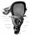
File:Lisser1911 fig39.jpg ==Fig. 39 Wax model of laryngeal region in human Embryo 22== [[:Category:Carnegie Embryo 22|'''Embryo no. 22''']] (20 mm.). (drawn from below)(2,340 × 2,746 (375 KB)) - 21:36, 15 June 2016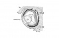
File:Streeter1957 fig06-20.jpg ...er1957 fig06-19.jpg|[[Carnegie stage 19]] ([[:Category:Carnegie Embryo 409|Embryo 409]]) ...er1957 fig06-20.jpg|[[Carnegie stage 20]] ([[:Category:Carnegie Embryo 462|Embryo 462]])(1,280 × 834 (133 KB)) - 21:14, 15 March 2017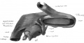
File:Lisser1911 fig37.jpg ==Fig. 37 Wax model of laryngeal region in human Embryo 22== [[:Category:Carnegie Embryo 22|'''Embryo no. 22''']] (20 mm.). (seen from below)(3,354 × 1,729 (349 KB)) - 21:22, 15 June 2016


