Search results
From Embryology
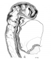
File:Wen1928-Fig04.jpg ==Fig. 4. Head of the 17-somite embryo H951== From the medial view of a model of the right side prepared at 150 diameters and reduced in the illustration to 100 diameters. The dotted line(1,200 × 1,392 (323 KB)) - 15:30, 21 April 2016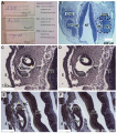
File:Human CS13-15 otic vesicle 01.jpg ==Human Otic Vesicle Carnegie Stages 13 to 15== Scale bar = 100 μm (B) and 150 μm (C-F).(1,574 × 1,779 (364 KB)) - 12:31, 6 April 2018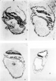
File:MartinFalkiner1938 plate02.jpg 6 Section 30 E. X 150. The amnion and embryonic plate are much damaged but the latter is now sect 7 Section 3.1. D. X 150. The embryonic plate is now cut through at 90°. The amnion has rapidly dim(1,280 × 1,840 (247 KB)) - 12:56, 11 August 2017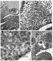
File:Crowder1957 plate01.jpg Fig. 8. Transverse section of embryo from beginning of 5th week: midgastric level. Aorta with origin of a mesone Fig. 9. From a section taken ahout 150 microns caudal to that of figure 8. Primordiunl diFlerentiated from surroun(1,280 × 1,457 (548 KB)) - 12:47, 27 May 2019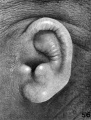
File:Streeter1922-fig56.jpg ==Fig. 56. Embryo No. 1782, 135.6 mm CRL== ...phs are all shown at an enlargement of 4 diameters. Specimens are from the Carnegie Collection, and length given is crown-rump.(592 × 783 (92 KB)) - 08:32, 28 January 2013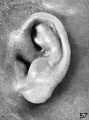
File:Streeter1922-fig57.jpg ==Fig. 57. Embryo No. 1702, 150 mm CRL== ...phs are all shown at an enlargement of 4 diameters. Specimens are from the Carnegie Collection, and length given is crown-rump.(577 × 775 (88 KB)) - 13:55, 22 November 2017
File:National Museum of Health and Medicine.jpg ...entre Collections]] that include the historic [[Carnegie Collection]] of [[Carnegie Embryos|human embryos]]. ...Developmental Anatomy Centre Collections]] | [[Carnegie_Collection]] | [[Carnegie Embryos]](800 × 600 (201 KB)) - 11:52, 19 December 2018


