Search results
From Embryology
Page title matches
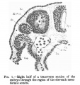
File:Low 01.jpg {{Low 1908}}(502 × 535 (60 KB)) - 02:44, 29 May 2017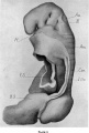
File:Low plate 01.jpg {{Low 1908}}(661 × 981 (137 KB)) - 00:11, 21 February 2012


Page text matches
- ...12]] | [[:File:Low 13.jpg|Fig 13]] | [[:File:Low 14.jpg|Fig 14]] | [[:File:Low 15.jpg|Fig 15]]952 bytes (124 words) - 03:45, 29 May 2017
- ...fetal tongue 01.jpg|image - fetal]] | [[:File:Human embryonic-fetal tongue 01.jpg|image - embryo and fetal]] | [[Tongue Development]] | [[Sensory_-_Taste_Dev937 bytes (124 words) - 15:00, 23 March 2016
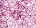
File:Mesentery histology 02.jpg ...stology 02.jpg|Low power view (x10)]] see also [[:File:Mesentery histology 01.jpg|High power (x40)]].(1,280 × 1,024 (256 KB)) - 13:49, 9 March 2018- ...e mount villi]] | [[:File:HillH52 chorionic villi 03.jpg|Chorion and villi low power]] | [[:File:HillH52 chorionic villi 04.jpg|Villi high power]] | [[:Fi638 bytes (78 words) - 21:54, 11 August 2014
File:Mouse lung development 03.jpg ...d high power)]] | [[:File:Mouse lung development 02.jpg|Normal mouse lung (low power)]] | [[Respiratory System Development]] | [[Mouse Development]] ...mage, resized to fit screen, reference label added) Mouse lung development 01.jpg(540 × 1,200 (349 KB)) - 11:27, 31 August 2017
File:Worm - embryonic cell lineage 02.jpg ...ulston, E. Schierenberg, J. G. White, J. N. Thomson. This image provides a low resolution overview of every cell in the developing c. elegans. :Links: [[:File:Worm - embryonic cell lineage 01.jpg|large full size image]] | [http://www.wormatlas.org/celllineages.html Worm(1,000 × 129 (19 KB)) - 05:51, 7 November 2010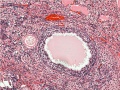
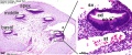
File:Ovary- atretic follicle 01.jpg ...y- atretic follicle 02.jpg|small image]] | [[:File:Ovary- atretic follicle 01.jpg|Large image]] | [[Ovary Development]] low magnification x10, UNSW Anatomy slide collection(793 × 595 (225 KB)) - 11:48, 9 May 2013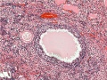
File:Ovary- atretic follicle 02.jpg ...y- atretic follicle 02.jpg|small image]] | [[:File:Ovary- atretic follicle 01.jpg|Large image]] | [[Ovary Development]] low magnification x10 , scaled to 600px UNSW Anatomy slide collection(600 × 450 (139 KB)) - 11:47, 9 May 2013- ...s.jpg|image - high power label]] | [[:File:Ovary histology 004.jpg|image - low power]] | [[:File:Ovary_histology_001.jpg|image - high power]] | [[:File:Ov1 KB (152 words) - 10:14, 6 May 2019
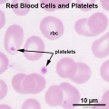
File:Platelet 01.jpg * Platelets are circulating fragments formed from a [[:File:Megakaryocyte 01.jpg|megakaryocyte]], a cell located only in the bone marrow. ** low platelet count (below 150,000) may lead to excessive bleeding.(600 × 600 (57 KB)) - 06:43, 7 March 2015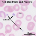
File:Platelet 02.jpg * Platelets are circulating fragments formed from a [[:File:Megakaryocyte 01.jpg|megakaryocyte]], a cell located only in the bone marrow. ** low platelet count (below 150,000) may lead to excessive bleeding.(600 × 600 (57 KB)) - 06:46, 7 March 2015- ! Low Power view | [[File:Mesentery histology 01.jpg|400px]]3 KB (355 words) - 13:48, 9 March 2018
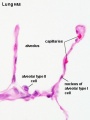
File:Respiratory histology 03.jpg [[File:Alveolar-sac-01.jpg|thumb|300px|Alveous cartoon]] ...apillary embedded in wall. Compare the structure with [[:File:Alveolar-sac-01.jpg|alveoli cartoon]].(450 × 600 (29 KB)) - 11:55, 8 April 2013- ...l Tube Defects|[[Abnormal Development - Folic Acid and Neural Tube Defects|Low Folic Acid]] File:Rubella virus 01.jpg|link=Abnormal Development - Viral Infection|[[Abnormal Development - Viral2 KB (241 words) - 09:53, 3 May 2020
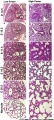
File:Human fetal cochlea 02.jpg Scale bars = 100 μm (low magnification) or 50 μm (high magnification). ...-_Inner_Ear_Development|'''Cochlea Links''']]: [[:File:Human fetal cochlea 01.jpg|Image week 8]] | [[:File:Human fetal cochlea 02.jpg|Image week 10]] | [[:Fi(1,270 × 532 (271 KB)) - 21:06, 12 May 2019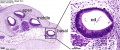
File:Human fetal cochlea 01.jpg Scale bars = 100 μm (low magnification) or 50 μm (high magnification). ...-_Inner_Ear_Development|'''Cochlea Links''']]: [[:File:Human fetal cochlea 01.jpg|Image week 8]] | [[:File:Human fetal cochlea 02.jpg|Image week 10]] | [[:Fi(1,270 × 532 (266 KB)) - 21:05, 12 May 2019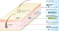
File:Touch receptors in mammalian skin cartoon.jpg ** include nociceptors and low-threshold C-fibers (Seal et al., 2009). :'''Links:''' [[:File:Touch receptors in mammalian skin cartoon 01.jpg|Image - Touch receptors 1]] | [[:File:Touch receptors in mammalian skin ca(800 × 429 (57 KB)) - 10:04, 25 March 2012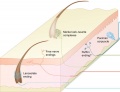
File:Touch receptors in mammalian skin cartoon 01.jpg ** include nociceptors and low-threshold C-fibers (Seal et al., 2009). :'''Links:''' [[:File:Touch receptors in mammalian skin cartoon 01.jpg|Image - Touch receptors 1]] | [[:File:Touch receptors in mammalian skin ca(600 × 459 (38 KB)) - 10:06, 25 March 2012- * Nasal Bridge - low [[File:Choanal atresia computed tomography 01.jpg|thumb|Choanal atresia computed tomography<ref name="PMID21772853"><pubmed>22 KB (331 words) - 23:11, 2 October 2012
- * Nasal Bridge - low [[File:Choanal atresia computed tomography 01.jpg|thumb|Choanal atresia computed tomography<ref name="PMID21772853"><pubmed>22 KB (329 words) - 14:08, 5 October 2011


