Search results
From Embryology
Page title matches
- ...gy_01.jpg|Palatine Tonsil]] | [[:File:Tonsil_histology_02.jpg|Tonsil]] | [[Immune System Development]]738 bytes (83 words) - 13:22, 24 February 2016
Page text matches
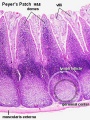
File:Peyer's patch 01.jpg ...o fold cells) - Function to transport gut lumen organisms and particles to immune cells across the epithelial barrier. {{Immune Images 2}}(450 × 600 (118 KB)) - 11:09, 16 January 2015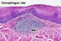
File:Oesophagus MALT.jpg {{Immune Images 2}} [[Category:Immune]] [[Category:Histology]] [[Category:Gastrointestinal Tract]](500 × 333 (73 KB)) - 11:44, 26 January 2015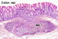
File:Colon MALT.jpg {{Immune Images 2}} [[Category:Immune]] [[Category:Histology]] [[Category:Gastrointestinal Tract]](500 × 333 (67 KB)) - 11:59, 26 January 2015
File:Tonsil histology 02.jpg {{Immune Images 2}} [[Category:Immune]] [[Category:Histology]] [[Category:Gastrointestinal Tract]](450 × 600 (62 KB)) - 10:46, 24 February 2012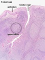
File:Tonsil histology 01.jpg # intimate contact between immune response effector cells {{Immune Images 2}}(450 × 600 (106 KB)) - 08:10, 18 February 2019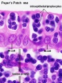
File:Peyer's patch 02.jpg ** function to transport gut lumen organisms and particles to immune cells across the epithelial barrier. {{Immune Images 2}}(450 × 600 (69 KB)) - 12:14, 26 January 2015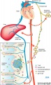
File:Gastrointestinal tract intestine immune cartoon 01.jpg ==Gastrointestinal Tract Immune== ...tem interaction through Mesenteric Lymph Nodes (MNLs) with the circulating immune system.(728 × 1,200 (284 KB)) - 11:47, 18 February 2019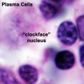
File:Plasma cell clockface nucleus 01.jpg {{Immune Images 2}} [[Category:Immune]] [[Category:Gastrointestinal Tract]](400 × 400 (27 KB)) - 10:36, 26 January 2015- ...B lymphocyte]] | [[:File:B lymphocytes EM08-10.jpg|B lymphocytes TEM]] | [[Immune System Development]] | [[Cardiovascular_System_-_Blood_Development|Blood]] [[Category:Immune]] [[Category:Blood]]1 KB (169 words) - 05:30, 26 January 2015
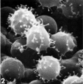
File:Lymphocyte rosettes EM02.jpg Fig. 2. SRBC showing microspherulation, with multiple microprojections with beaded {{lymphocyte images}}(661 × 665 (62 KB)) - 16:14, 17 February 2019
File:Intraepithelial lymphocyte differentiation 02.jpg {{Immune Images 2}} Type Of Use: post on a website Figure 2.(1,200 × 557 (79 KB)) - 14:18, 26 January 2015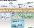
File:Intraepithelial lymphocyte differentiation 03.jpg {{Immune Images 2}} [[Category:Immune]] [[Category:Gastrointestinal Tract]][[Category:Cartoon]](600 × 484 (68 KB)) - 14:56, 26 January 2015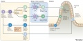
File:Intraepithelial lymphocyte differentiation 01.jpg {{Immune Images 2}} [[Category:Immune]] [[Category:Gastrointestinal Tract]][[Category:Cartoon]](1,200 × 585 (108 KB)) - 18:26, 30 April 2018- File:T_and_B_lymphocytes_EM10.jpg|T and B Lymphocytes 2 TEM File:B lymphocyte EM09.jpg|B lymphocyte 2 TEM2 KB (314 words) - 06:39, 13 January 2016
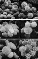
File:Lymphocyte rosettes EM01-06.jpg Fig. 2. SRBC showing microspherulation, with multiple microprojections with beaded {{lymphocyte images}}(1,364 × 2,100 (334 KB)) - 16:14, 17 February 2019- | '''Week 2''' [https://moodle.telt.unsw.edu.au/mod/book/view.php?id=789982&chapterid=1 ...w.edu.au/mod/book/view.php?id=789982&chapterid=100772 Lymphatic Tissue and Immune System] | '''Week 8''' [https://moodle.telt.unsw.edu.au/mod/book/view.php?i15 KB (2,032 words) - 15:10, 7 June 2019
- Development of the immune system will also link to cardiovascular development notes (blood and vessel {{Immune Links}}7 KB (1,062 words) - 10:25, 18 February 2019
- **Special:NewFiles|New images ** Week 2|Week 23 KB (334 words) - 16:01, 22 June 2019
- # [[Special:NewFiles|New images]] ====Week 2====8 KB (981 words) - 00:33, 31 July 2018
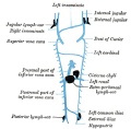
File:Gray0592.jpg ...ppear, at the junction of the subclavian vein with the primitive jugular; (2) posterior sac, at the junction of the iliac vein with the cardinal; (3) re {{Grays Lymphatic images}}(600 × 590 (55 KB)) - 12:49, 15 February 2013


