Search results
From Embryology
Page title matches
- ...[Book_-_Manual_of_Human_Embryology_18-5|Blood-vascular System in Series of Human Embryos]] | [[Book_-_Manual_of_Human_Embryology_18-6|Arteries]] | [[Book_-_1 KB (155 words) - 14:29, 14 January 2017
- ...[Book_-_Manual_of_Human_Embryology_18-5|Blood-vascular System in Series of Human Embryos]] | [[Book_-_Manual_of_Human_Embryology_18-6|Arteries]] | [[Book_-_1 KB (154 words) - 11:32, 15 December 2016
- 854 bytes (114 words) - 15:45, 16 January 2015
- ...[Book_-_Manual_of_Human_Embryology_18-5|Blood-vascular System in Series of Human Embryos]] | [[Book_-_Manual_of_Human_Embryology_18-6|Arteries]] | [[Book_-_1 KB (151 words) - 18:16, 14 January 2017
- 370 bytes (44 words) - 18:39, 17 August 2017
- 27 bytes (3 words) - 22:46, 31 March 2014
- :[[Book - Manual of Human Embryology 18-2|'''Development of the Heart''']]: [[:File:Keibel_Mall_2_372.jpg|Fig. 372. S446 bytes (59 words) - 12:30, 3 September 2015
- {{Human Embryology Manual 2 18}} [[File:Julius Tandler.jpg|thumb|alt=Julius Tandler|link=Embryology History - Julius Tandler|Julius Tandler (1869 – 1936)]]88 KB (14,592 words) - 10:33, 16 September 2020
- File:Keibel_Mall_2_373.jpg|Fig. 373. — Model of the heart of a human embryo. No. 300 of Rob. Meyer's collection ...eibel_Mall_2_378.jpg|Fig. 378. — Model of the heart of the embryo Hah of 5.2 mm. greatest length. In the collection of the I.4 KB (567 words) - 00:50, 2 April 2014
Page text matches
File:Keibel Mall 2 370.jpg ==Fig. 370. Blood-cells from a hepatic vessel of a human embryo of 11 mm== {{Human Embryology Manual 2 18-1}}(1,000 × 312 (40 KB)) - 11:08, 29 March 2014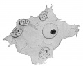
File:Keibel Mall 2 360.jpg {{Human Embryology Manual 2 18-1}} {{Human Embryology Manual 2 18}}(995 × 800 (110 KB)) - 10:59, 29 March 2014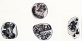
File:Keibel Mall 2 365.jpg ==Fig. 365. Degenerating human leucocytes== {{Human Embryology Manual 2 18-1}}(1,000 × 503 (75 KB)) - 11:05, 29 March 2014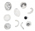
File:Keibel Mall 2 369.jpg ==Fig. 369. — Blood-corpuscles from the vessels of a human fetus of eight months== {{Human Embryology Manual 2 18-1}}(793 × 700 (55 KB)) - 11:07, 29 March 2014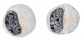
File:Keibel Mall 2 363.jpg {{Human Embryology Manual 2 18-1}} {{Human Embryology Manual 2 18}}(900 × 436 (69 KB)) - 11:02, 29 March 2014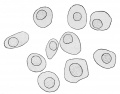
File:Keibel Mall 2 367.jpg ==Fig. 367. Outlines of erythrocytes of a human embryo of 8 mm== {{Human Embryology Manual 2 18-1}}(1,021 × 800 (76 KB)) - 11:06, 29 March 2014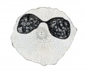
File:Keibel Mall 2 364.jpg {{Human Embryology Manual 2 18-1}} {{Human Embryology Manual 2 18}}(800 × 673 (78 KB)) - 11:03, 29 March 2014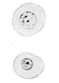
File:Keibel Mall 2 355.jpg ==Fig. 355. Two blood-corpuscles of a human embryo of 4 mm== {{Human Embryology Manual 2 18-1}}(564 × 800 (35 KB)) - 10:56, 29 March 2014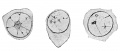
File:Keibel Mall 2 354.jpg ==Fig. 354. Three primitive mesamoeboids from the yolk-sac of a human embryo of about 1 mm== {{Human Embryology Manual 2 18-1}}(1,000 × 429 (48 KB)) - 10:54, 29 March 2014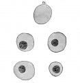
File:Keibel Mall 2 358.jpg ==Fig. 358. Four blood-corpuscles from a human embryo of 15.5 mm== {{Human Embryology Manual 2 18-1}}(786 × 806 (57 KB)) - 10:57, 29 March 2014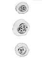
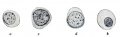
File:Keibel Mall 2 356.jpg ==Fig. 356. — Three blood-corpuscles of a human embryo of 7.5 mm== {{Human Embryology Manual 2 18-1}}(584 × 800 (35 KB)) - 10:56, 29 March 2014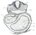
File:Keibel Mall 2 376.jpg {{Human Embryology Manual 2 18-2}} {{Human Embryology Manual 2 18}}(1,000 × 985 (172 KB)) - 11:13, 29 March 2014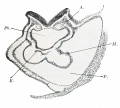
File:Keibel Mall 2 372.jpg {{Human Embryology Manual 2 18-2}} {{Human Embryology Manual 2 18}}(998 × 900 (112 KB)) - 19:10, 23 June 2019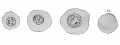
File:Keibel Mall 2 357.jpg ==Fig. 357. Three blood-corpuscles from a human embryo of 9.4 mm== {{Human Embryology Manual 2 18-1}}(1,000 × 379 (46 KB)) - 10:56, 29 March 2014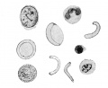
File:Keibel Mall 2 359.jpg ==Fig. 359. Blood-corpuscles from a blood-vessel of a human embryo of eight months== {{Human Embryology Manual 2 18-1}}(854 × 700 (50 KB)) - 10:58, 29 March 2014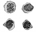
File:Keibel Mall 2 362.jpg ==Fig. 362. Four small lymphocytes from normal human blood== {{Human Embryology Manual 2 18-1}}(674 × 600 (52 KB)) - 11:02, 29 March 2014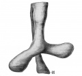
File:Keibel Mall 2 341.jpg {{Human Embryology Manual 2 17-10}} {{Human Embryology Manual 2 17}}(865 × 800 (65 KB)) - 11:43, 29 July 2019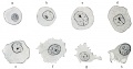
File:Keibel Mall 2 361.jpg ==Fig. 361. Red blood-cells from the placental chorion of a human embryo of 15 mm== {{Human Embryology Manual 2 18-1}}(1,000 × 522 (80 KB)) - 11:00, 29 March 2014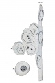
File:Keibel Mall 2 368.jpg .... 368. — Endothelium and blood-cells from the lower part of the aorta of a human embryo of 9.4 mm== {{Human Embryology Manual 2 18-1}}(664 × 1,000 (71 KB)) - 11:06, 29 March 2014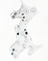
File:Keibel Mall 2 371.jpg ==Fig. 371. Hepatic cylinders of a human embryo of 11 mm== {{Human Embryology Manual 2 18-1}}(785 × 1,000 (89 KB)) - 11:55, 16 January 2016


