Search results
From Embryology
Page title matches
- [[Fetal Development|fetus]]<noinclude>[[Category:Template]][[Category:Term Link]][[Category:Fetal]][[168 bytes (18 words) - 09:48, 20 March 2020
- 272 members (50 subcategories, 140 files) - 09:37, 10 October 2017

File:Ultrasound Fetus 01.mp4 (3.45 MB) - 16:40, 12 March 2013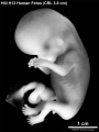
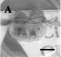
File:HillH13 Fetus bf01.jpg ==Human Fetus (Week 9)== HillH13 fetus week 9 CRL 3.8 cm left view.(1,200 × 1,600 (171 KB)) - 08:41, 14 December 2014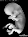
File:HillH13 Fetus bf02.jpg ==Human Fetus (Week 9)== HillH13 fetus week 9 CRL 3.8 cm left view.(1,200 × 1,600 (170 KB)) - 08:40, 14 December 2014File:Ultrasound Fetus 02.mp4 (782 KB) - 16:12, 16 March 2013- ...hin the humerus by bone marrow as an arbitrary definition of the embryo to fetus transition. ...recognized in a given specimen, that specimen is straightway classed as a fetus.''"694 bytes (95 words) - 13:04, 13 June 2019
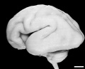
File:Human Fetus CRL240mm brain.jpg ==Human Fetus Brain== [[Category:Human Fetus]][[Category:Neural]](1,280 × 1,030 (129 KB)) - 16:05, 27 March 2018
File:Dog fetus day 30.jpg ==Dog Fetus day 28==(400 × 375 (32 KB)) - 16:39, 25 September 2012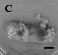
File:Dog fetus day 32.jpg ==Dog Fetus day 28==(400 × 375 (28 KB)) - 16:40, 25 September 2012- ===Fetus (12 weeks)=== Movie shows a 12 week fetus in 3d in realtime (hence 4D).765 bytes (103 words) - 09:36, 19 May 2015
- ===Fetus (19 weeks)=== Movie shows a 19 week fetus in 3 dimensional (3D) in realtime (hence 4D).805 bytes (111 words) - 00:48, 18 June 2014
File:Ultrasound Fetus 01-icon.jpg (659 × 480 (39 KB)) - 16:40, 12 March 2013- | valign="top" |Movie shows a 12 week fetus in 3d in realtime (hence 4D). The beginning of the movies shows the ventral (anterior) view of the fetus head to top.781 bytes (110 words) - 16:14, 16 March 2013
- | valign="top" |Movie shows a 19 week fetus in 3 dimensional (3D) in realtime (hence 4D). The beginning of the movies shows the fetus lying with head to right and lower limbs to left. The overlay shows region703 bytes (105 words) - 16:38, 12 March 2013
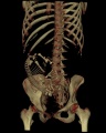
File:Fetus 35 week CT.jpg [[Category:Human Fetus]] [[Category:Computed Tomography]](700 × 874 (62 KB)) - 14:21, 24 August 2010- 60 bytes (12 words) - 21:25, 4 April 2010
File:Dog fetus day 28.jpg ==Dog Fetus day 28==(400 × 375 (24 KB)) - 16:38, 25 September 2012
File:Human week 10 fetus 03.jpg ==Human Female Fetus - Pelvic Region (10 week)== {{Human Female Fetus Week 10 gallery}}(1,600 × 1,200 (370 KB)) - 17:21, 25 May 2016
File:Human week 10 fetus 23.jpg ==Human Female Fetus - Pelvic Region (10 week)== {{Human Female Fetus Week 10 gallery}}(1,600 × 1,200 (393 KB)) - 17:21, 25 May 2016


Page text matches
- <gallery caption="Human Female Fetus (week 10)"> File:Human week 10 fetus 01.jpg|Sagittal Section (plane D)733 bytes (105 words) - 11:57, 30 May 2016
- ...of the Fetus 15|15 Nutrition and Metabolism]] | [[Book - Physiology of the Fetus 16|Figures]]<noinclude>[[Category:Template]][[Category:Reference]][[Categor1 KB (150 words) - 09:44, 10 September 2018
- ...through Uterus and Fetus at Birth]] | [[:File:BrauneC2.jpg|Uterus without Fetus at Birth]] | [[Embryology_History_-_17th_and_18th_Century_Anatomies#Wilhelm544 bytes (76 words) - 16:09, 2 November 2016
- File:BrauneB1.jpg|Uterus and Fetus Position at Term File:BrauneB2.jpg|Section through Uterus and Fetus305 bytes (48 words) - 10:05, 10 November 2012

File:Koala fetus.jpg ==Koala Fetus== Koala fetus near birth.(559 × 1,000 (77 KB)) - 16:32, 1 December 2010File:HillH13 Fetus bf01.jpg ==Human Fetus (Week 9)== HillH13 fetus week 9 CRL 3.8 cm left view.(1,200 × 1,600 (171 KB)) - 08:41, 14 December 2014File:HillH13 Fetus bf02.jpg ==Human Fetus (Week 9)== HillH13 fetus week 9 CRL 3.8 cm left view.(1,200 × 1,600 (170 KB)) - 08:40, 14 December 2014- ===Fetus (12 weeks)=== Movie shows a 12 week fetus in 3d in realtime (hence 4D).765 bytes (103 words) - 09:36, 19 May 2015
- ...m]] | [[:File:Low1909 fig06.jpg| fetus 55 mm]] | [[:File:Low1909 fig07.jpg|fetus 95 mm]] | [[:File:Low1909 plate01.jpg|human 18-24-95 mm]] |438 bytes (63 words) - 15:12, 6 January 2017
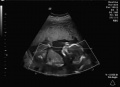
File:Ultrasound12wk 3D image.jpg ...ultrasound static image of the 12 week fetus shows a ventral view with the fetus upside down, with the head down and cord to the top. ...ategory:Genetic Abnormalities]] [[Category:Human Embryo]] [[Category:Human Fetus]](301 × 248 (8 KB)) - 15:10, 11 October 2009- ===Fetus (19 weeks)=== Movie shows a 19 week fetus in 3 dimensional (3D) in realtime (hence 4D).805 bytes (111 words) - 00:48, 18 June 2014
File:HillH13 Fetus.gif ==Human Fetus (Week 9)== HillH13 fetus week 9 CRL 3.8 cm left view. (stereo pair animated gif)(450 × 600 (274 KB)) - 08:40, 14 December 2014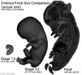
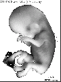
File:Size comparison embryo-fetus actual.jpg ==Size Comparison of the Embryo to Fetus== Original file name: EFsizeactual.jpg (Size comparison embryo-fetus actual.jpg)(194 × 178 (14 KB)) - 17:45, 6 May 2011
File:Lineback1920 fig06-7.jpg ===Fig. 6. Cross-section of the ascending colon of a human fetus=== Cross-section of the ascending colon of a human fetus 105 mm. CR. length, showing the three tseniaj in a triangular position in t(1,200 × 808 (177 KB)) - 09:13, 17 January 2013- The 29-day fetus . . . . . . . . . . . . . . . . . . . . . . . . . . . . . . . . . . . . . . The 31-day fetus . . . . . . . . . . . . . . . . . . . . . . . . . . . . . . . . . . . . . .2 KB (49 words) - 13:51, 4 May 2018
- Fetus week 9-10 9-10 Week Fetus 1530 bytes (84 words) - 17:55, 12 August 2011
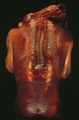
File:Spina Bifida 1.jpg ==Spina Bifida Fetus== Dorsal view of fetus with extensive spina bifida and anencephaly.(393 × 599 (49 KB)) - 10:28, 11 May 2016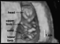
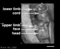
File:Ultrasound12wk 3D image2.jpg ==Ultrasound Fetus (12 week)== Image from near end of movie showing ventral view of fetus head to top, upper limbs, lower limbs and umbilical cord visible.(362 × 264 (8 KB)) - 09:06, 6 November 2012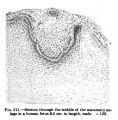
File:Keibel Mall 211.jpg ==Fig. 211 Human Fetus Integumentary Gland==(427 × 436 (43 KB)) - 13:01, 24 August 2012
File:Human week 10 fetus 02.jpg ==Human Female Fetus (10 week)== See also [[:File:Human week 10 fetus 01.jpg|'''Large Image Version''']](800 × 462 (83 KB)) - 14:38, 11 October 2015


