Search results
From Embryology
Page title matches
- 62 bytes (6 words) - 09:24, 17 April 2018
- ...x|left]] This 1957 paper by Smith describes development of the trachea and esophagus and includes several embryos from the [[Carnegie Collection]]. Note different spelling USA {{esophagus}} or UK {{oesophagus}}.61 KB (9,187 words) - 14:29, 5 May 2019
- A CASE OF NORMAL EMBRYONIC ATRESIA OF THE ESOPHAGUS for a study of the history of the primordial germ cells, the esophagus9 KB (1,364 words) - 15:46, 30 September 2020
- ...is 1917 historic paper by Jordan describes normal embryonic atresia of the esophagus. =A Case of Normal Embryonic Atresia of the Esophagus=9 KB (1,379 words) - 09:10, 2 October 2020
- =A Case of Atresia of the Esophagus Combined with Traoheoesophageal Fistula in a 9 mm Human Embryo, and its Emb ...the most frequent malformations of esophagus and trachea is atresia of the esophagus combined with tracheocsophageal fistula. The great number and the almost id18 KB (2,910 words) - 15:51, 2 April 2017
Page text matches

File:Smith1957 fig01.jpg ==Fig. 1. Vogt’s classification of atresia of the esophagus== Type I, atresia of entire esophagus.(1,000 × 583 (73 KB)) - 11:13, 24 August 2016- ...y:Reference]][[Category:Historic Embryology]][[Category:1910's]][[Category:Esophagus]][[Category:Abnormal Development]]</noinclude>427 bytes (54 words) - 15:30, 30 September 2020
- ...rly development of the trachea and esophagus in relation to atresia of the esophagus and tracheoesophageal fistula''']]. (1957) Contributions To Embryology, No.365 bytes (46 words) - 12:51, 3 May 2020
- Esophagus ...egut derivatives (other than pharynx, lower respiratory tract, most of the esophagus) are supplied by the celiac trunk.416 bytes (59 words) - 12:44, 23 January 2019
- ...sis: II. Tension of differential growth as a stimulus to myogenesis in the esophagus''']]. (1920) J Gen Physiol. 20;3(1): 61-83. [https://www.ncbi.nlm.nih.gov/p541 bytes (70 words) - 18:18, 11 August 2017
- | Esophageal atresia || Foregut ( Esophagus || occurs in 8th week || 1 in 3000-4500 birth- results from deviation of tr ...oregut ( Esophagus || during week 8 || Due to incomplete recanalisation of Esophagus1 KB (140 words) - 15:50, 19 October 2014
- | Esophageal atresia || Foregut ( Esophagus || occurs in 8th week || 1 in 3000-4500 birth- results from deviation of tr ...oregut ( Esophagus || during week 8 || Due to incomplete recanalisation of Esophagus1 KB (139 words) - 15:03, 19 October 2014
- ...uman embryo, and its embryological explanation|'''A case of atresia of the esophagus combined with traoheoesophageal fistula in a 9 mm human embryo, and its emb467 bytes (59 words) - 18:40, 27 May 2019
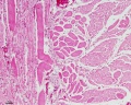
File:Smooth muscle histology 006.jpg * human esophagus(1,280 × 1,024 (481 KB)) - 14:21, 23 February 2013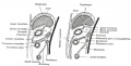
File:Gray0990.jpg ...mall sac lying within the thorax on the right side of the lower end of the esophagus. The anterior layer of the transverse mesocolon is at first distinct from t(800 × 407 (60 KB)) - 15:56, 28 April 2011- ...the absence of severe malformations. There is no ideal replacement for the esophagus and the optimal surgical treatment for patients with long-gap EA is still c2 KB (244 words) - 17:09, 9 May 2015
File:Embryo stage 22 C7L.jpg * relative postion of esophagus and trachea(619 × 389 (67 KB)) - 11:33, 31 May 2010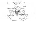
File:Lisser1911 fig01.jpg ...cricoarytacnoideus posterior. V. C., vertebral column; Esoph. L., Lumen of esophagus; constr., constrictor muscle; cri. post, M. cricoarytaenoide'uspostcior; n.(1,119 × 900 (92 KB)) - 14:50, 12 June 2016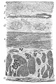
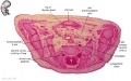
File:Gray1033.jpg ==Fig. 1033. Section of the Human Esophagus==(399 × 600 (96 KB)) - 12:09, 11 May 2014- ...is 1917 historic paper by Jordan describes normal embryonic atresia of the esophagus. =A Case of Normal Embryonic Atresia of the Esophagus=9 KB (1,379 words) - 09:10, 2 October 2020

File:Human week 10 fetus 08.jpg ..., epiglottis, arytenoid cartilage, arytænoideus muscle, cricoid cartilage, esophagus, trachea, vertebra(1,200 × 900 (323 KB)) - 21:03, 8 October 2015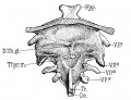
File:Sudler1902-fig05.jpg D. Th. g1., ductus thyreoglossus; Hyp., hypophysis; 0e., (esophagus; Tr., trachea; V. P.’ V. P.’’, V. P./” and V. PJV, flrst, second,(1,000 × 769 (182 KB)) - 15:37, 24 May 2017
File:Gray0621.jpg ...on of the seventh rib with its cartilage; (b) middle, to the glands on the esophagus and to those around the termination of the inferior vena cava; and (c) post ...and lateral aortic glands and to the glands on the terminal portion of the esophagus.(599 × 800 (131 KB)) - 13:00, 15 February 2013- A CASE OF NORMAL EMBRYONIC ATRESIA OF THE ESOPHAGUS for a study of the history of the primordial germ cells, the esophagus9 KB (1,364 words) - 15:46, 30 September 2020
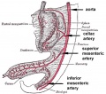
File:GIT blood supply.jpg # '''Foregut''' - celiac artery (Adult: pharynx, esophagus, stomach, upper duodenum, respiratory tract, liver, gallbladder pancreas)(568 × 500 (47 KB)) - 12:48, 17 April 2019


