Search results
From Embryology
Page title matches
- This {{Embryology}} category shows content related to the {{cranial nerve}} development (cerebral nerves) that are associated with the brain and the # {{CN I}} – Olfactory nerve74 members (0 subcategories, 33 files) - 14:54, 26 May 2018
- ...]]<noinclude>[[Category:Template]][[Category:Term Link]][[Category:Cranial Nerve]][[Category:Ectoderm]][[Category:Sensory]][[Category:Neural]]</noinclude>204 bytes (23 words) - 12:35, 6 May 2018
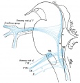
- ...Ciliary ganglion originating from neural crest of caudal diencephalon and cranial mesencephalon ...igeminal ganglion originating from neural crest of caudal diencephalon and cranial mesencephalon; trigeminal placode935 bytes (111 words) - 14:03, 22 June 2015
- There are twelve pairs of cranial nerves; they are attached to the brain and are transmitted through foramina {{Cranial Nerve Table collapsible}}956 bytes (136 words) - 11:48, 6 February 2016
- ! colspan="2"|[[Neural - Cranial Nerve Development|Cranial Nerves]] |}<noinclude>[[Category:Cranial Nerve]][[Category:Table]][[Category:Template]]</noinclude>608 bytes (79 words) - 12:49, 6 May 2018
- ...} | {{CN X}} | {{CN XI}} | {{CN XII}} | {{placodes}} | [[:Category:Cranial Nerve]] ! [[Historic_Embryology_Papers|'''Historic Embryology''']] Cranial Nerves 1 KB (176 words) - 11:59, 30 October 2018
- ! colspan="5"|[[Neural - Cranial Nerve Development|Cranial Nerves]] ! Nerve Number2 KB (212 words) - 13:01, 6 May 2018
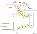
- ==Cranial Nerves== ...aryngeal placodes) give rise to the distal cells of the sensory ganglia of cranial nerves V, VII, IX and X.4 KB (633 words) - 11:47, 29 March 2016
- ...pg|thumb|Human Embryo CNS ([[Carnegie stage 14|stage 14]]) showing cranial nerve development]] [[File:Neural - cranial nerves.jpg|thumb|Cranial nerves]]21 KB (2,950 words) - 05:30, 10 December 2019
- ...:Term Link]][[Category:Neural]][[Category:Neural Crest]][[Category:Cranial Nerve]]</noinclude>207 bytes (25 words) - 15:02, 27 June 2018
- MeSH Terms: Cranial Nerves/embryology ===Cranial Pair 0: The Nervus Terminalis===18 KB (2,570 words) - 13:28, 12 May 2019
File:Mouse cranial nerve model SHH.jpg ==Mouse Cranial Nerve Model - Sonic Hedgehog== ...om rhombomere 2 (r2) or 4 (r4) interact with placodal cells to develop the cranial nerves.(954 × 900 (74 KB)) - 14:03, 6 May 2018- ...om rhombomere 2 (r2) or 4 (r4) interact with placodal cells to develop the cranial nerves Cranial nerve development requires co-ordinated Shh and canonical Wnt signaling.2 KB (283 words) - 18:28, 14 April 2016
- <center>See also [[Neural - Cranial Nerve Development]]</center> {{Cranial Nerve Links}}14 KB (1,910 words) - 13:46, 19 February 2019
- ===Cranial Pair 0: The Nervus Terminalis=== ...t knowledge on this structure, incorporating original illustrations of the nerve at different developmental stages, and focuses on its anatomical and clinic2 KB (281 words) - 13:29, 12 May 2019
- {{Cranial Nerve Table collapsible}} ...ths]] | [[Neural Exam - 18 Cranial Nerves|18 months]] | [[Neural - Cranial Nerve Development]]1 KB (166 words) - 08:37, 16 February 2016

Page text matches
File:Gray0698.jpg ==Fig. 698. Primary Terminal Nuclei of the Afferent (sensory) Cranial Nerves== * Cranial Nerve 8 (CNVIII) to cochlear nuclei (hearing) and vestibular nuclei (balance)(500 × 518 (47 KB)) - 04:34, 27 April 2014- {{Cranial Nerve Table collapsible}} ...ths]] | [[Neural Exam - 18 Cranial Nerves|18 months]] | [[Neural - Cranial Nerve Development]]730 bytes (95 words) - 08:38, 16 February 2016
File:Gray0697.jpg ==Fig. 697. Cranial motor nerves brainstem nuclei of origin== * Cranial nerve 7 (CNVII) facial nerve to facial motor nucleus(500 × 540 (49 KB)) - 04:32, 27 April 2014- ...]]<noinclude>[[Category:Template]][[Category:Term Link]][[Category:Cranial Nerve]][[Category:Ectoderm]][[Category:Sensory]][[Category:Neural]]</noinclude>204 bytes (23 words) - 12:35, 6 May 2018
- This {{Embryology}} category shows content related to the {{cranial nerve}} development (cerebral nerves) that are associated with the brain and the # {{CN I}} – Olfactory nerve74 members (0 subcategories, 33 files) - 14:54, 26 May 2018

File:Human week 10 fetus 12.jpg ==Human Female Fetus - Olfactory Nerve (10 week)== ...ed from the nasal placode, the olfactory receptor neurons form an afferent nerve fibre transmitting the [[Sensory - Smell Development|sense of smell]] to ol(1,200 × 900 (349 KB)) - 14:38, 25 May 2016- ...:Term Link]][[Category:Neural]][[Category:Neural Crest]][[Category:Cranial Nerve]]</noinclude>207 bytes (25 words) - 15:02, 27 June 2018
- {{Cranial Nerve Table collapsible}} ...ths]] | [[Neural Exam - 18 Cranial Nerves|18 months]] | [[Neural - Cranial Nerve Development]]1,021 bytes (145 words) - 08:38, 16 February 2016
- {{Cranial Nerve Table collapsible}} ...ths]] | [[Neural Exam - 18 Cranial Nerves|18 months]] | [[Neural - Cranial Nerve Development]]1 KB (166 words) - 08:37, 16 February 2016
- ...p" |[[File:Newborn n 02.jpg|right|150px]]Examination of the baby’s cranial nerve function is often accomplished by observing spontaneous activity. * During crying, facial movement ('''CN VII''') Cranial Nerve 7 is observed for fullness or asymmetry.2 KB (245 words) - 08:37, 16 February 2016
- ! colspan="2"|[[Neural - Cranial Nerve Development|Cranial Nerves]] |}<noinclude>[[Category:Cranial Nerve]][[Category:Table]][[Category:Template]]</noinclude>608 bytes (79 words) - 12:49, 6 May 2018
File:Mouse cranial nerve model SHH.jpg ==Mouse Cranial Nerve Model - Sonic Hedgehog== ...om rhombomere 2 (r2) or 4 (r4) interact with placodal cells to develop the cranial nerves.(954 × 900 (74 KB)) - 14:03, 6 May 2018- ...toric Embryology]][[Category:1800's]][[Category:Neural]][[Category:Cranial Nerve]]</noinclude>304 bytes (39 words) - 14:36, 4 December 2019
- ...} | {{CN X}} | {{CN XI}} | {{CN XII}} | {{placodes}} | [[:Category:Cranial Nerve]] ! [[Historic_Embryology_Papers|'''Historic Embryology''']] Cranial Nerves 1 KB (176 words) - 11:59, 30 October 2018
- {{Cranial Nerve Table collapsible}} ...ths]] | [[Neural Exam - 18 Cranial Nerves|18 months]] | [[Neural - Cranial Nerve Development]]1 KB (174 words) - 08:38, 16 February 2016
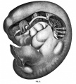
File:Mall1905 fig3.jpg ...reconstructed; the tenth and twelfth cranial nerves and the flrst cervical nerve by [[Embryology History - George Streeter|Dr. Streeter]]. [[Category:Cranial Nerve]](902 × 1,000 (155 KB)) - 12:00, 17 February 2016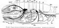
File:Bailey368.jpg ==Fig. 368. Diagram showing the distribution of the cranial nerves in the Amniota== [[Category:Cranial Nerve]](1,074 × 523 (134 KB)) - 08:20, 16 February 2016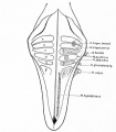
File:Bailey390.jpg ...point of attachment of the acoustic and the sensory root of the trigeminal nerve is shown by dotted circles; the motor nuclei are represented by heavy dots. [[Category:Cranial Nerve]](583 × 667 (50 KB)) - 23:01, 4 September 2014- ...nce]][[Category:Historic Embryology]][[Category:1800's]][[Category:Cranial Nerve]]</noinclude>370 bytes (43 words) - 12:24, 7 April 2020
- ...0 paper by Landacre described development of the sensory components of the cranial ganglia. '''Modern Notes:''' {{cranial nerve}} | {{neural}}430 bytes (56 words) - 09:30, 23 February 2020


