File:Morphology of Heart Tube Formation- Student Image .png
From Embryology

Size of this preview: 463 × 600 pixels. Other resolution: 681 × 882 pixels.
Original file (681 × 882 pixels, file size: 283 KB, MIME type: image/png)
Original student image based off: [1]
Description
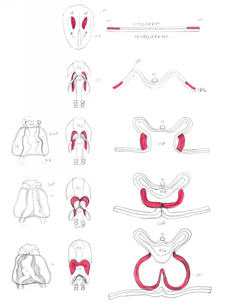
Figure 2
(Anterior, ventral, cross-section views)
HF- Heart Fields shown in red to show the fusion and folding of splanchnic mesoderm, CR- Cardiogenic Region, CC- Cardiac Crescent, AIP- Anterior Intestinal Portal, VV- Vitelline Vein, EHT- Endocardial Heart Tube, HT- Heart Tube, PC- Pericardial cavity, FG- Foregut, SoF- Site of Fusion, V- Ventricle, M- Myocardium, N- Notochord, SPL- Splanchnopleure, SM- Splanchnic Mesoderm
File history
Yi efo/eka'e gwa ebo wo le nyangagi wuncin ye kamina wunga tinya nan
| Gwalagizhi | Nyangagi | Dimensions | User | Comment | |
|---|---|---|---|---|---|
| current | 22:14, 17 September 2017 | | 681 × 882 (283 KB) | Z5076019 (talk | contribs) | Original student image based off: [http://dev.biologists.org/content/144/13/2381.figures-only] |
You cannot overwrite this file.
File usage
The following 2 pages use this file: