File:Week6.jpg
From Embryology
Size of this preview: 545 × 599 pixels. Other resolution: 621 × 683 pixels.
Original file (621 × 683 pixels, file size: 89 KB, MIME type: image/jpeg)
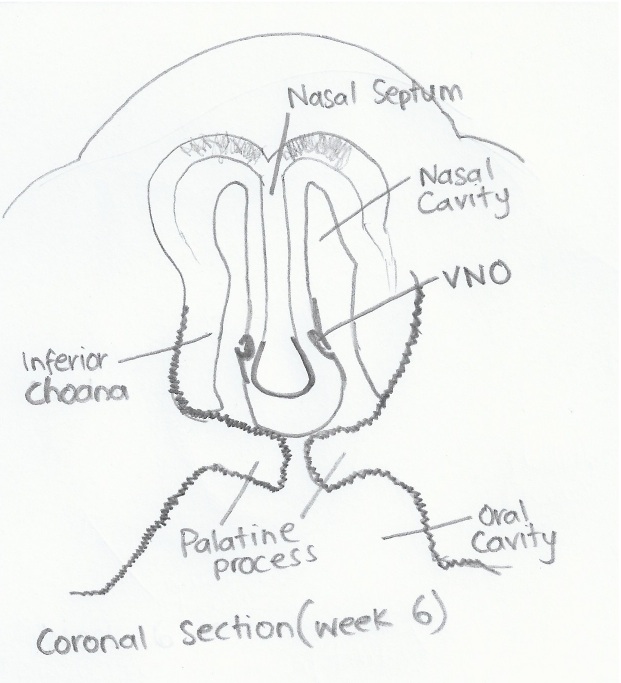
A diagram of the coronal section of an embryo at week 6 of development, indicating the formation of the vomeronasal organ, choana and palatine processes.
Image is self drawn by Student based on histology provided by: <pubmed>15454774</pubmed>
File history
Click on a date/time to view the file as it appeared at that time.
| Date/Time | Thumbnail | Dimensions | User | Comment | |
|---|---|---|---|---|---|
| current | 02:43, 3 October 2012 | | 621 × 683 (89 KB) | Z3331264 (talk | contribs) |
You cannot overwrite this file.
File usage
The following 2 pages use this file: