Uploads by Z8600021
From Embryology
This special page shows all uploaded files.
| Date | Name | Thumbnail | Size | Description | Versions |
|---|---|---|---|---|---|
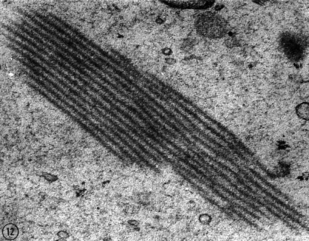
| 00:30, 24 February 2012 | Human pronuclear stage EM12.jpg (file) |  |
265 KB | 1 | |
| 00:29, 24 February 2012 | Human pronuclear stage EM11.jpg (file) |  |
130 KB | 1 | |
| 00:20, 24 February 2012 | Human pronuclear stage EM10.jpg (file) |  |
140 KB | 1 | |
| 00:20, 24 February 2012 | Human pronuclear stage EM08.jpg (file) |  |
56 KB | ==Human Pronuclear Stage Endoplasmic Reticulum== * Large endoplasmic reticulum vesicles (er) in budding activity. * The buds have tubular proflies and are provided with saccular terminations which are similar to the small ER vesicles scattered in the oo | 1 |
| 00:16, 24 February 2012 | Human pronuclear stage EM09.jpg (file) |  |
53 KB | ==Human Pronuclear Stage Endoplasmic Reticulum== * Large endoplasmic reticulum vesicles (er) in budding activity. * The buds have tubular proflies and are provided with saccular terminations which are similar to the small ER vesicles scattered in the oo | 1 |
| 00:10, 24 February 2012 | Human pronuclear stage EM07.jpg (file) |  |
52 KB | ==Human Pronuclear Stage Endoplasmic Reticulum== * Large endoplasmic reticulum vesicles (er) in budding activity. * The buds have tubular proflies and are provided with saccular terminations which are similar to the small ER vesicles scattered in the oo | 1 |
| 00:06, 24 February 2012 | Human pronuclear stage EM06.jpg (file) |  |
245 KB | ==Human Pronuclear Stage Mitochondria== * Region of the ooplasm of the penetrated ovum with typical complement of organelles. * The mitochondria are numerous and mostly spheroidal. * Two mitochondria with archlike arrangement of the inner cristae are i | 1 |
| 00:00, 24 February 2012 | Human pronuclear stage EM03-05.jpg (file) |  |
104 KB | ==Human Pronuclear Stage Mitochondria== Fig. 3. Spheroidal mitochondria represent the most common type of mitochondria to be encountered in the ovum (see also Fig. 5). They have limited numbers of cristae which are arranged roughly parallel to the outer | 1 |
| 23:46, 23 February 2012 | Human pronuclear stage EM022.jpg (file) |  |
225 KB | ==Human Pronuclear Stage== * Region of the penetrated human ovum with male and female pronuclei (PN). * The pronuclear material appears to be highly hydrated, although zonally condensed in patchy aggregates. * Nucleoli (n) and intrapronuclear annulate | 1 |
| 23:45, 23 February 2012 | Human pronuclear stage EM02.jpg (file) |  |
194 KB | ==Human Pronuclear Stage== * Region of the penetrated human ovum with male and female pronuclei (PN). * The pronuclear material appears to be highly hydrated, although zonally condensed in patchy aggregates. * Nucleoli (n) and intrapronuclear annulate | 1 |
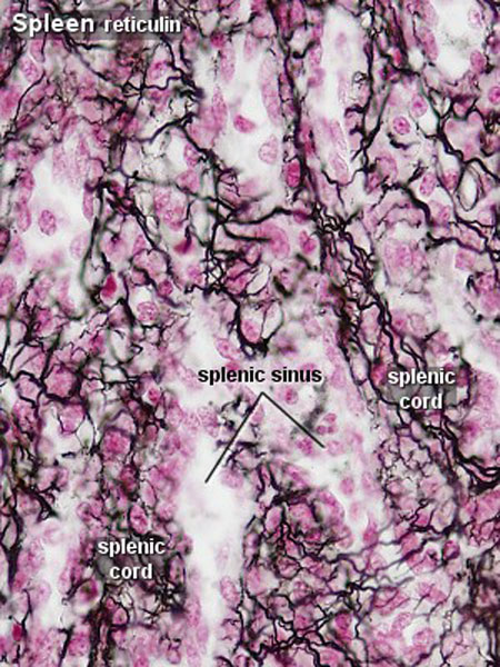
| 19:41, 23 February 2012 | Spleen histology 04.jpg (file) |  |
108 KB | 2 | |
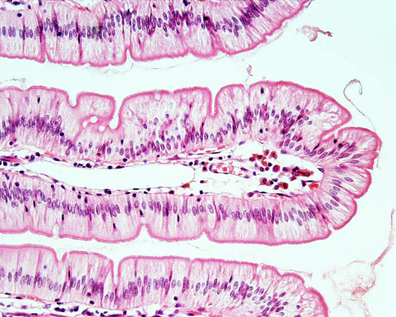
| 16:52, 23 February 2012 | Intestine histology 002.jpg (file) |  |
130 KB | 2 | |
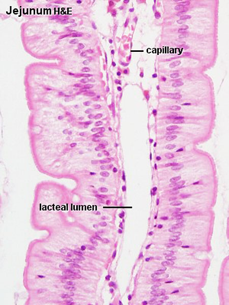
| 16:42, 23 February 2012 | Intestine histology 001.jpg (file) |  |
65 KB | Jej20he.jpg | 1 |
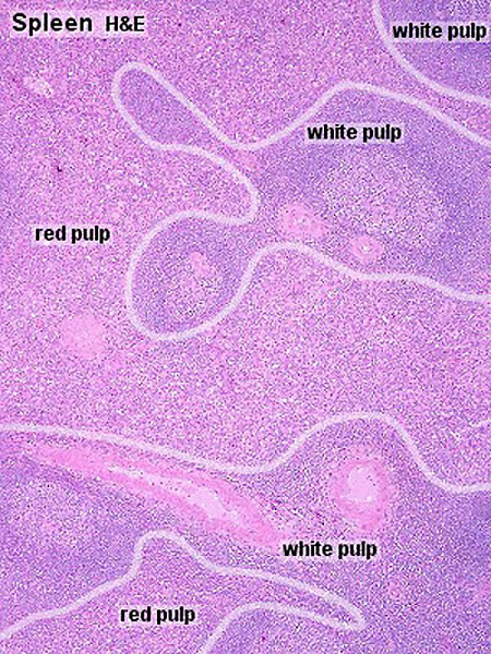
| 20:30, 22 February 2012 | Spleen histology 03.jpg (file) |  |
83 KB | 2 | |
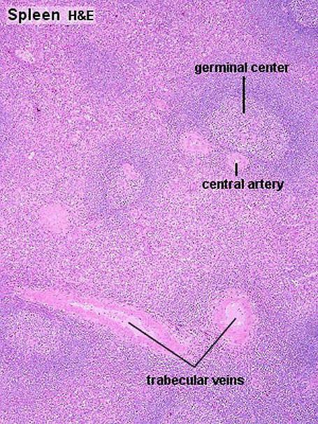
| 20:27, 22 February 2012 | Spleen histology 02.jpg (file) |  |
142 KB | 2 | |
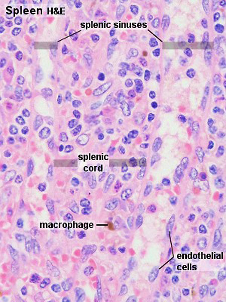
| 20:26, 22 February 2012 | Spleen histology 01.jpg (file) |  |
133 KB | 2 | |
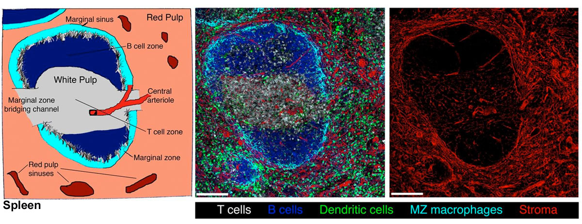
| 19:55, 22 February 2012 | Spleen structure 02.jpg (file) |  |
46 KB | 1 | |
| 19:55, 22 February 2012 | Spleen structure 01.jpg (file) |  |
211 KB | 1 | |
| 19:53, 22 February 2012 | Lymph node structure 02.jpg (file) |  |
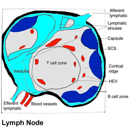
54 KB | ==Lymph node structure== Schematic representation of the organization of a lymph node (left panel). Afferent lymphatics enter lymph nodes and deliver lymph to the subcapsular sinus (SCS), which forms a channel around the periphery of the lymph node. Lymp | 1 |
| 19:48, 22 February 2012 | Lymph node structure 01.jpg (file) |  |
167 KB | ==Lymph node structure== Schematic representation of the organization of a lymph node (left panel). Afferent lymphatics enter lymph nodes and deliver lymph to the subcapsular sinus (SCS), which forms a channel around the periphery of the lymph node. Lymp | 1 |
| 19:15, 22 February 2012 | Lymph node model dymamics.jpg (file) |  |
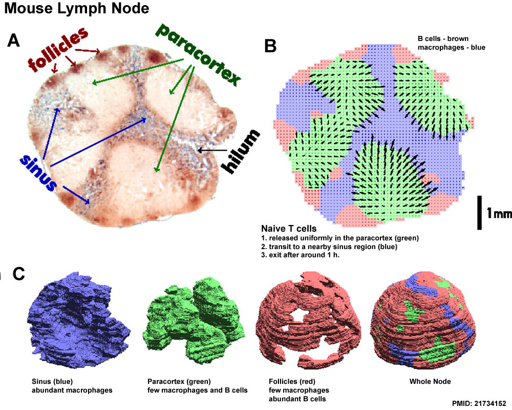
231 KB | ==Mouse Lymph Node Dynamics== Modeling the naive T cell LN transit. (A) Microtome slice from a mesenteric rat LN with B cells stained in brown and macrophages stained in blue. (B) Using image processing algorithms (SI Text), the lymph node volume was div | 1 |
| 17:30, 22 February 2012 | T2 lymphocyte EM14.jpg (file) |  |
156 KB | ==T2 lymphocyte Electron Micrograph== Fig. 14. Lymph node cells (4 days after immunization with DBA/2 mastocytoma) labeled with aBA0 followed by SaRIg-Pox. This cell classified as T2 lymphocyte is characterized by its large content in polyribosomes, ofte | 1 |
| 17:23, 22 February 2012 | T2 lymphocyte EM13.jpg (file) |  |
177 KB | ==T2 lymphocyte Electron Micrograph== FIg. 13. Spleen cell (10 days after alloantigenic immunization with DBA/2 mastocytoma) labeled with aMSLA detected by the bridge technique with phage T4. Some of the phage heads are sectioned tangentially and theref | 1 |
| 16:49, 22 February 2012 | Plasma cell EM06.jpg (file) |  |
84 KB | ==Plasma Cell Electron Micrograph== Fig 6. Plasma cells labeled by incubations with aMBLA, followed by the "bridge"technique with SaRIg-Pox. Plasma cells, especially when fully mature, showed also a reduced amount of MBLA compared with B lymphocytes. | 1 |
| 16:11, 22 February 2012 | T and B lymphocytes EM10.jpg (file) |  |
155 KB | 1 | |
| 16:11, 22 February 2012 | T and B lymphocytes EM09.jpg (file) |  |
137 KB | T and B_lymphocytes_EM09.jpg | 1 |
| 14:39, 22 February 2012 | B lymphocyte EM10.jpg (file) |  |
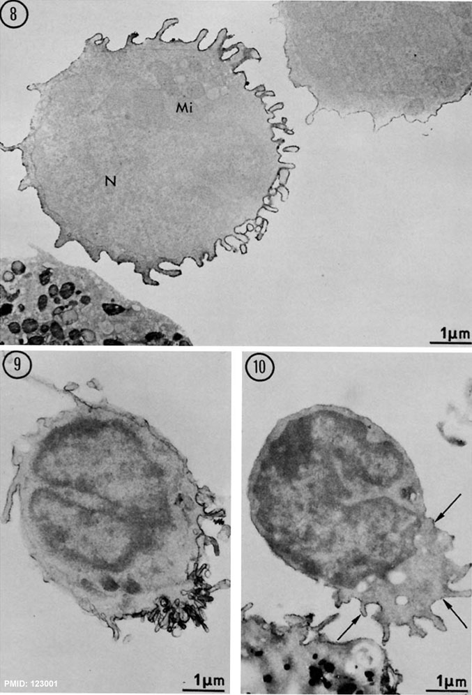
92 KB | ==Human B Lymphocyte Electron Micrograph== Fig. 10. Uropod-bearing lymphocyte exhibiting a "hand-mirror profile". sIg are detected by anti-Ig antibodies on the villous processes of the posterior end of this cell (arrows). A neighboring monocyte is also l | 1 |
| 14:37, 22 February 2012 | B lymphocyte EM09.jpg (file) |  |
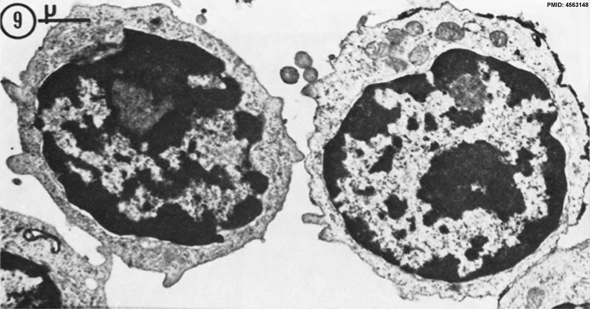
89 KB | ==Human B Lymphocyte Electron Micrograph== Fig. 9. This IgM-bearing lymphocyte exhibits a more complete polar concentration of labeled microvilli. The remaining surface is poorly labeled. x 12,000. {{lymphocyte images}} ===References=== <pubmed> | 1 |
| 14:36, 22 February 2012 | B lymphocyte EM08.jpg (file) |  |
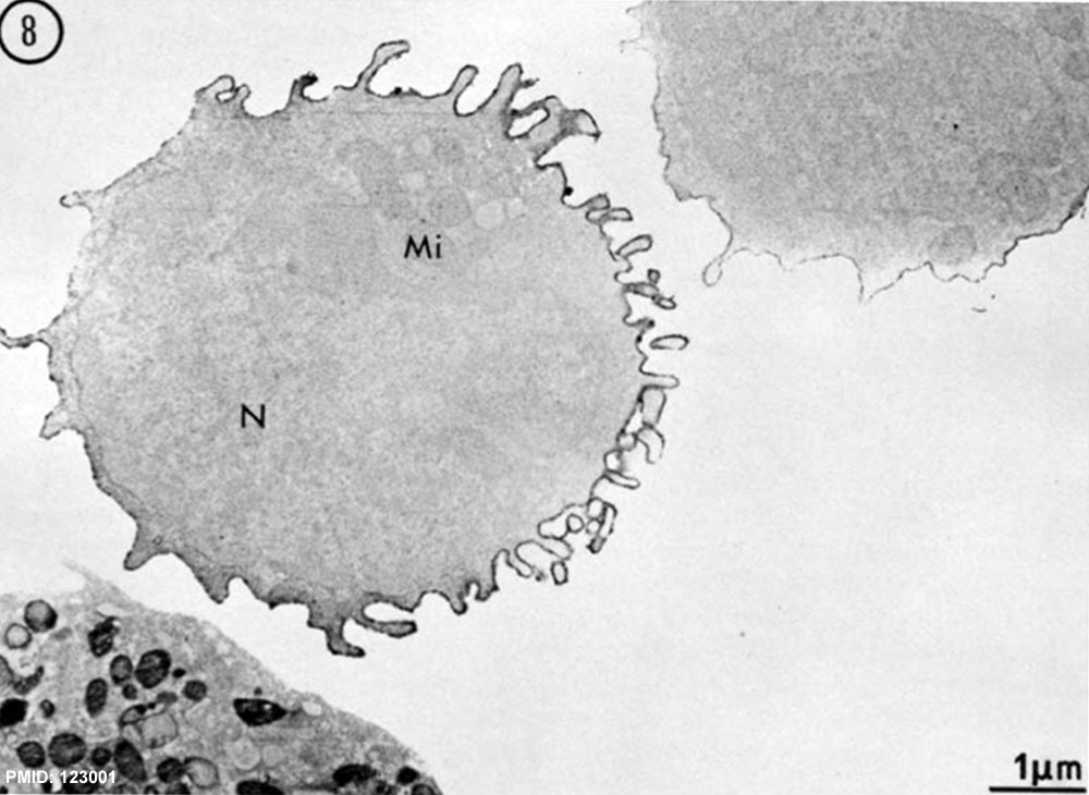
111 KB | ==Human B Lymphocyte Electron Micrographs== Fig. 8 Normal B lymphocyte with surface IgM. Microvilli are flowing toward one pole of the cell, the remaining membrane being relatively smoother and moderately labeled. An intermediate B lymphocyte is seen at | 1 |
| 14:35, 22 February 2012 | B lymphocytes EM08-10.jpg (file) |  |
124 KB | ==Human B Lymphocyte Electron Micrographs== Fig. Normal B lymphocyte with surface IgM. Microvilli are flowing toward one pole of the cell, the remaining membrane being relatively smoother and moderately labeled. An intermediate B lymphocyte is seen at up | 1 |
| 13:56, 22 February 2012 | Lymphocyte rosettes EM012.jpg (file) |  |
59 KB | 1 | |
| 13:56, 22 February 2012 | Lymphocyte rosettes EM06.jpg (file) |  |
66 KB | 1 | |
| 13:55, 22 February 2012 | Lymphocyte rosettes EM05.jpg (file) |  |
55 KB | 1 | |
| 13:55, 22 February 2012 | Lymphocyte rosettes EM04.jpg (file) |  |
53 KB | 1 | |
| 13:55, 22 February 2012 | Lymphocyte rosettes EM03.jpg (file) |  |
51 KB | 1 | |
| 13:55, 22 February 2012 | Lymphocyte rosettes EM02.jpg (file) |  |
62 KB | 1 | |
| 13:54, 22 February 2012 | Lymphocyte rosettes EM01.jpg (file) |  |
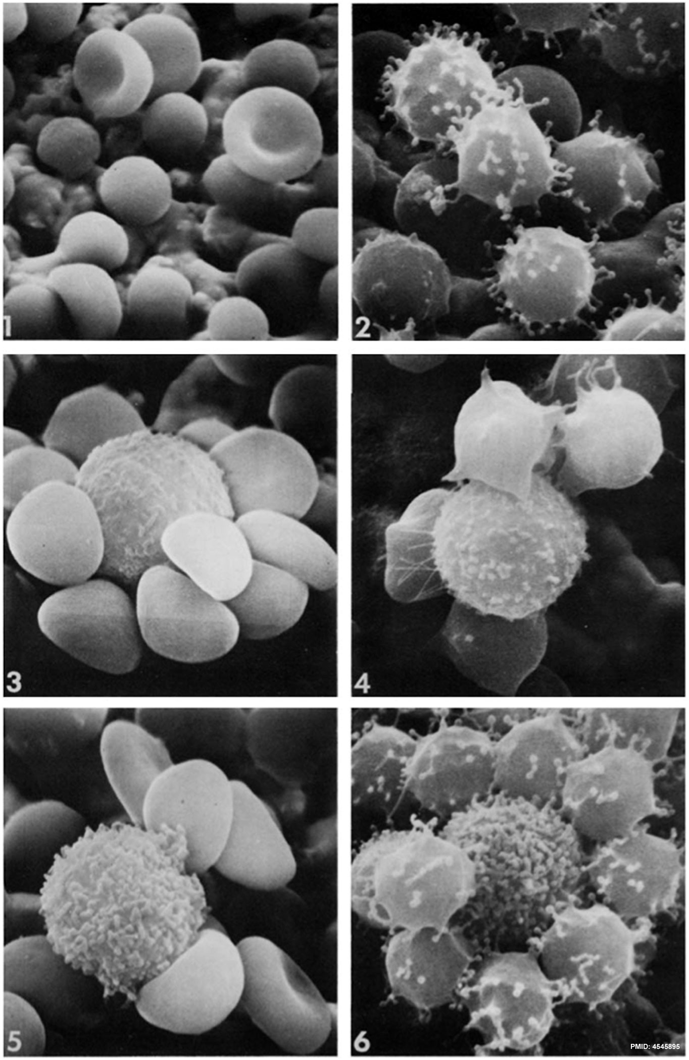
58 KB | ==Human Lymphocytes with Sheep Erythrocyte Rosettes== Human lymphocytes (Thymocytes, T-cells) and Sheep erythrocytes (Red Blood Cells, SRBC). Fig. 1. SRBC washed with HBSS showing spheroechinocyte transformation. Two SRBC have maintained their biconcav | 1 |
| 13:47, 22 February 2012 | Lymphocyte rosettes EM01-06.jpg (file) |  |
334 KB | Sheep Red Blood Cells Fig. 1. SRBC washed with HBSS showing spheroechinocyte transformation. Two SRBC have maintained their biconcaveshape. X 3,600. Fig. 2. SRBC showing microspherulation, with multiple microprojections with beaded ends. X 6,900. Fig. | 1 |
| 17:07, 21 February 2012 | Carnegie stage 23 OPT.jpg (file) |  |
36 KB | 1 | |
| 17:07, 21 February 2012 | Carnegie stage 22 OPT.jpg (file) |  |
38 KB | 1 | |
| 17:07, 21 February 2012 | Carnegie stage 21 OPT.jpg (file) |  |
39 KB | 1 | |
| 17:06, 21 February 2012 | Carnegie stage 20 OPT.jpg (file) |  |
47 KB | 1 | |
| 17:06, 21 February 2012 | Carnegie stage 19 OPT.jpg (file) |  |
46 KB | 1 | |
| 17:05, 21 February 2012 | Carnegie stage 18 OPT.jpg (file) |  |
43 KB | 1 | |
| 17:05, 21 February 2012 | Carnegie stage 17 OPT.jpg (file) |  |
57 KB | 1 | |
| 17:04, 21 February 2012 | Carnegie stage 16 OPT.jpg (file) |  |
51 KB | ==Carnegie Stage 16 Optical Projection Tomography Model== Still image of the left lateral side of 3 dimensional OPT models of human embryo. * developmental stage - CS16 * specimen number - N519 * karyotype - 46XY Embryo cropped and resized from figur | 1 |
| 17:03, 21 February 2012 | Carnegie stage 15 OPT.jpg (file) |  |
56 KB | ==Carnegie Stage 15 Optical Projection Tomography Model== Still image of the left lateral side of 3 dimensional OPT models of human embryo. * developmental stage - CS15 * specimen number - N340 * karyotype - 46XX Embryo cropped and resized from figur | 1 |
| 17:02, 21 February 2012 | Carnegie stage 14 OPT.jpg (file) |  |
55 KB | ==Carnegie Stage 13 Optical Projection Tomography Model== Still image of the left lateral side of 3 dimensional OPT models of human embryo. * developmental stage - CS13 * specimen number - N383 * karyotype - 46XY Carnegie_stage_13_OPT.jpg Embryo cropp | 1 |
| 17:02, 21 February 2012 | Carnegie stage 13 OPT.jpg (file) |  |
53 KB | ==Carnegie Stage 13 Optical Projection Tomography Model== Still image of the left lateral side of 3 dimensional OPT models of human embryo. * developmental stage - CS13 * specimen number - N383 * karyotype - 46XY Carnegie_stage_13_OPT.jpg Embryo cropp | 1 |
| 16:59, 21 February 2012 | Carnegie stage 12 OPT.jpg (file) |  |
46 KB | ==Carnegie Stage 12 Optical Projection Tomography Model== Still image of the left lateral side of 3 dimensional OPT models of human embryo. * developmental stage - CS12 * specimen number - N285 * karyotype - 46XX Embryo cropped and resized from figur | 1 |