File:Ultrasound4.jpg: Difference between revisions
From Embryology
(Amniocentesis in first trimester pregnant sheep. The 22 Gauge needle is being guided using a 3.5MHz ultrasound transducer. Picture courtesy of A David) |
No edit summary |
||
| Line 1: | Line 1: | ||
Amniocentesis in first trimester pregnant sheep. The 22 Gauge needle is being guided using a 3.5MHz ultrasound transducer. | Amniocentesis in first trimester pregnant sheep. The 22 Gauge needle is being guided using a 3.5MHz ultrasound transducer. | ||
''This image was reproduced with written permission from the author.'' | |||
Dr Anna David PhD MRCOGSenior Lecturer and Honorary Consultant in Obstetrics and Maternal/FetalMedicine, | |||
Institute for Women's Health, | |||
University College London and UCLH, | |||
86 - 96 Chenies Mews,London,WC1E 6HX | |||
Tel: +44-20-7679-6651 | |||
Fax: +44-7383-7429 | |||
Mob: +44-7852-220375 | |||
Latest revision as of 12:17, 23 September 2010
Amniocentesis in first trimester pregnant sheep. The 22 Gauge needle is being guided using a 3.5MHz ultrasound transducer.
This image was reproduced with written permission from the author.
Dr Anna David PhD MRCOGSenior Lecturer and Honorary Consultant in Obstetrics and Maternal/FetalMedicine,
Institute for Women's Health,
University College London and UCLH,
86 - 96 Chenies Mews,London,WC1E 6HX
Tel: +44-20-7679-6651
Fax: +44-7383-7429
Mob: +44-7852-220375
File history
Click on a date/time to view the file as it appeared at that time.
| Date/Time | Thumbnail | Dimensions | User | Comment | |
|---|---|---|---|---|---|
| current | 11:24, 10 September 2010 | 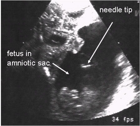 | 449 × 402 (56 KB) | Z3254753 (talk | contribs) | Amniocentesis in first trimester pregnant sheep. The 22 Gauge needle is being guided using a 3.5MHz ultrasound transducer. Picture courtesy of A David |
You cannot overwrite this file.
File usage
The following page uses this file: