File:Apoptotic trophoblast.jpg: Difference between revisions
(Student drawing (by Wyatt Ng, z5001524) This diagram illustrates the structure of the placenta and chorionic villi. During the formation of placenta, some trophoblast cells, including mostly syncytiotrophoblast and some erythroblast, will undergo apopt...) |
(Z5001524 uploaded a new version of "File:Apoptotic trophoblast.jpg") |
(No difference)
| |
Revision as of 21:11, 23 July 2014
Student drawing (by Wyatt Ng, z5001524) This diagram illustrates the structure of the placenta and chorionic villi. During the formation of placenta, some trophoblast cells, including mostly syncytiotrophoblast and some erythroblast, will undergo apoptosis and the DNA of the fetus will be fragmented. Eventually, they will be able to distribute and migrate into the maternal circulation through passing the intervillus space, which is filled with maternal blood
Copyright Statement: Beginning six months after publication, I z5001524 grant the public the non-exclusive right to copy, distribute, or display the Work under a Creative Commons Attribution-Noncommercial-Share Alike 3.0 Unported license, as described at http://creativecommons.org/licenses/by-nc-sa/3.0/ and http://creativecommons.org/licenses/by-nc-sa/3.0/legalcode.
File history
Yi efo/eka'e gwa ebo wo le nyangagi wuncin ye kamina wunga tinya nan
| Gwalagizhi | Nyangagi | Dimensions | User | Comment | |
|---|---|---|---|---|---|
| current | 21:18, 23 July 2014 | 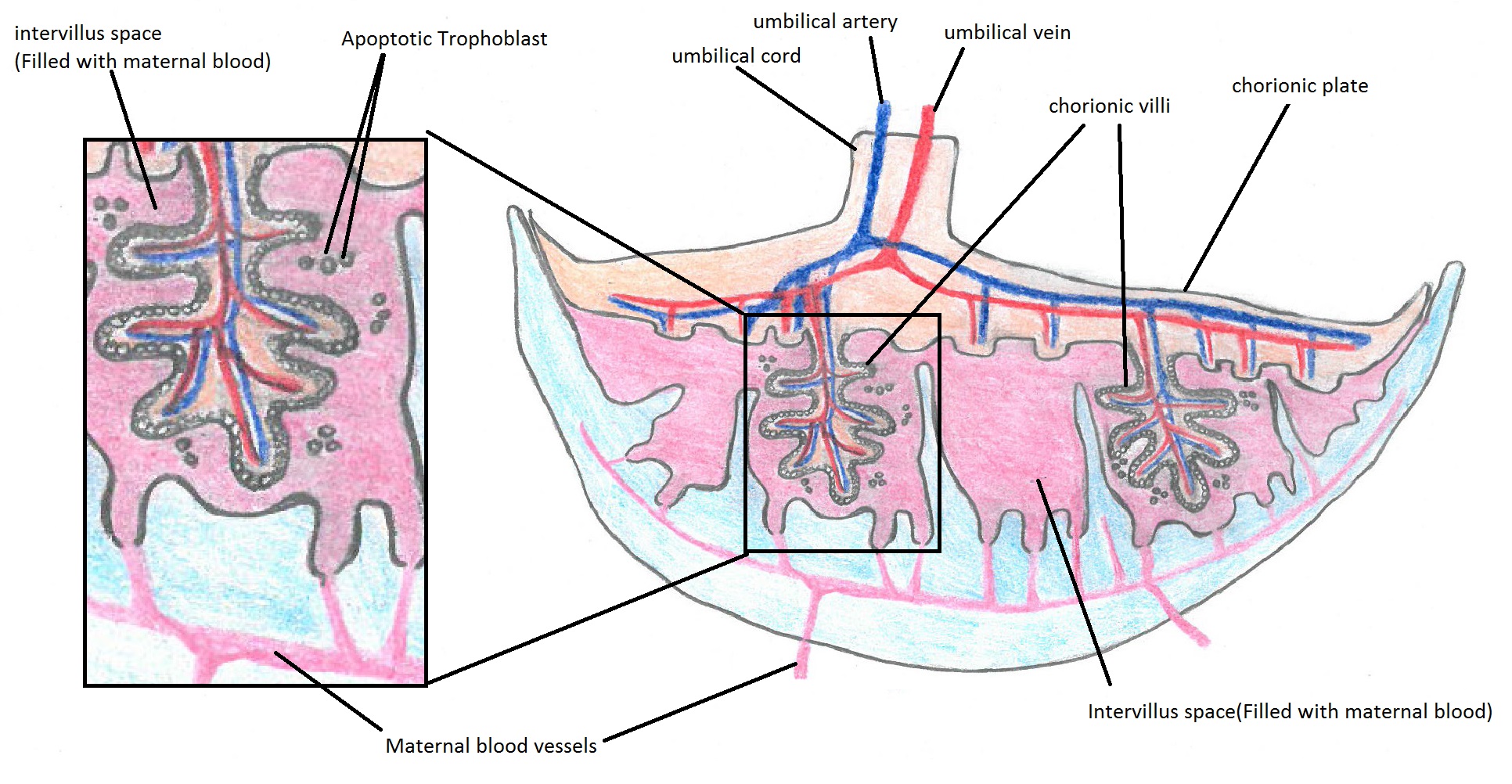 | 1,932 × 1,002 (517 KB) | Z5001524 (talk | contribs) | Reverted to version as of 09:11, 21 July 2014 |
| 21:14, 23 July 2014 | Error creating thumbnail: File with dimensions greater than 12.5 MP | 6,121 × 2,393 (2.76 MB) | Z5001524 (talk | contribs) | Modified drawing by adding utertine glands, decidua basalis and myometrium. | |
| 21:11, 23 July 2014 | Error creating thumbnail: File with dimensions greater than 12.5 MP | 6,121 × 2,393 (2.76 MB) | Z5001524 (talk | contribs) | Modified drawing by adding utertine glands, decidua basalis and myometrium. | |
| 19:11, 21 July 2014 |  | 1,932 × 1,002 (517 KB) | Z5001524 (talk | contribs) | Student drawing (by Wyatt Ng, z5001524) This diagram illustrates the structure of the placenta and chorionic villi. During the formation of placenta, some trophoblast cells, including mostly syncytiotrophoblast and some erythroblast, will undergo apopt... |
You cannot overwrite this file.
File usage
There are no pages that use this file.