File:Aortic valve disease.jpg: Difference between revisions
No edit summary |
No edit summary |
||
| Line 1: | Line 1: | ||
'''Loss of NOTCH1 expression in proximity to calcific nodules in human aortic valves.''' (A) Representative sections from control (A,B) and diseased (C-F) aortic valve cusps. (B) is high magnification image of boxed area in (A) and (D,E) are higher magnification of region in (C) while image in (F) shows another calcified aortic valve. Expression of Notch1 intracellular domain (NICD) is found in the thickened fibrosa of diseased aortic valve (C,E) as compared to the acellular fibrosa of control valves (A,B). However, there is significant loss of NICD expression in cells residing adjacent to calcific nodules (D,F). The fibrosa is oriented upward in all panels, and scale bars equal 100 microns (B,D,E,F are at same magnification). Brown signal represents NICD expression while nuclei are counterstained in blue.<ref name="PMID22110751" | '''Loss of NOTCH1 expression in proximity to calcific nodules in human aortic valves.''' (A) Representative sections from control (A,B) and diseased (C-F) aortic valve cusps. (B) is high magnification image of boxed area in (A) and (D,E) are higher magnification of region in (C) while image in (F) shows another calcified aortic valve. Expression of Notch1 intracellular domain (NICD) is found in the thickened fibrosa of diseased aortic valve (C,E) as compared to the acellular fibrosa of control valves (A,B). However, there is significant loss of NICD expression in cells residing adjacent to calcific nodules (D,F). The fibrosa is oriented upward in all panels, and scale bars equal 100 microns (B,D,E,F are at same magnification). Brown signal represents NICD expression while nuclei are counterstained in blue.<ref name="PMID22110751"><pubmed>22110751</pubmed></ref> | ||
[https://www.ncbi.nlm.nih.gov/pmc/articles/PMC3218038/| Direct link to full article] | [https://www.ncbi.nlm.nih.gov/pmc/articles/PMC3218038/| Direct link to full article] | ||
Revision as of 15:53, 26 October 2016
Loss of NOTCH1 expression in proximity to calcific nodules in human aortic valves. (A) Representative sections from control (A,B) and diseased (C-F) aortic valve cusps. (B) is high magnification image of boxed area in (A) and (D,E) are higher magnification of region in (C) while image in (F) shows another calcified aortic valve. Expression of Notch1 intracellular domain (NICD) is found in the thickened fibrosa of diseased aortic valve (C,E) as compared to the acellular fibrosa of control valves (A,B). However, there is significant loss of NICD expression in cells residing adjacent to calcific nodules (D,F). The fibrosa is oriented upward in all panels, and scale bars equal 100 microns (B,D,E,F are at same magnification). Brown signal represents NICD expression while nuclei are counterstained in blue.[1]
Copyright
This is an open-access article distributed under the terms of the Creative Commons Attribution License, which permits unrestricted use, distribution, and reproduction in any medium, provided the original author and source are credited.
- Note - This image was originally uploaded as part of an undergraduate science student project and may contain inaccuracies in either description or acknowledgements. Students have been advised in writing concerning the reuse of content and may accidentally have misunderstood the original terms of use. If image reuse on this non-commercial educational site infringes your existing copyright, please contact the site editor for immediate removal.
- ↑ <pubmed>22110751</pubmed>
File history
Click on a date/time to view the file as it appeared at that time.
| Date/Time | Thumbnail | Dimensions | User | Comment | |
|---|---|---|---|---|---|
| current | 15:49, 26 October 2016 | 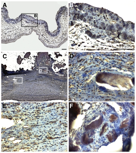 | 434 × 489 (241 KB) | Z3462474 (talk | contribs) | '''Loss of NOTCH1 expression in proximity to calcific nodules in human aortic valves.''' (A) Representative sections from control (A,B) and diseased (C-F) aortic valve cusps. (B) is high magnification image of boxed area in (A) and (D,E) are higher mag... |
You cannot overwrite this file.
File usage
The following 2 pages use this file: