Talk:2009 Lecture 21
From Embryology
Background Reading
- Chemokine signaling guides regional patterning of the first embryonic artery http://genesdev.cshlp.org/content/23/19/2272.abstract?etoc
- The aorta traverses the body, yet little is known about how it is patterned in different anatomical locations. Here, we show that the aorta develops from genetically distinct endothelial cells originating from diverse locations within the embryo. Furthermore, chemokine (C-X-C motif) receptor 4a (cxcr4a) is restricted to endothelial cells derived from anterior mesoderm, and is required specifically for formation of the lateral aortae. Cxcl12b, a cxcr4a ligand, is expressed in endoderm underlying the lateral aortae, and loss of cxcl12b phenocopies cxcr4a deficiency. These studies reveal unexpected endothelial diversity within the aorta that is necessary to facilitate its regional patterning by local cues.
- http://www.plosone.org/article/info%3Adoi%2F10.1371%2Fjournal.pone.0006267 Citation: Rochais F, Dandonneau M, Mesbah K, Jarry T, Mattei M-G, et al. (2009) Hes1 Is Expressed in the Second Heart Field and Is Required for Outflow Tract Development. PLoS ONE 4(7): e6267. doi:10.1371/journal.pone.0006267
- Tu C-T, Yang T-C, Tsai H-J (2009) Nkx2.7 and Nkx2.5 Function Redundantly and Are Required for Cardiac Morphogenesis of Zebrafish Embryos. PLoS ONE 4(1): e4249. doi:10.1371/journal.pone.0004249 http://www.plosone.org/article/info%3Adoi%2F10.1371%2Fjournal.pone.0004249
- Vascular system of the human spinal cord in the prenatal period: a dye injection and corrosion casting study
- "The vascularization of the spinal cord was investigated in 50 human fetuses aged from 10 to 28 gestational weeks using dye injection methods and corrosion casting accompanied by scanning electron microscopy. In the investigated period of fetal development, the general vascular architecture of the spinal cord, corresponding to that described postnatally, seemed to be already established. The observed changes included: (1) remodeling of the supplying (extrinsic) arterial branches, (2) transformation of the posterior anastomotic chain into two distinct posterior spinal arteries, and (3) development of the capillary networks in the gray and white matter."
- "The remodeling of the radicular arteries supplying the spinal cord was accompanied by a decrease in their number and transition from regular to irregular distribution (appearance of intersegmental differences in their frequency). The anterior spinal artery and regular array of the central arteries were already present in the youngest fetuses examined, but the final remodeling of the posterior anastomotic chain into two posterior spinal arteries occurred between 15th and 20th week of fetal life indicating that the vascularization of the anterior region of the spinal cord in the investigated period of fetal life was more advanced as compared with that of the posterior region."
- "The capillary network of the gray matter in the youngest fetuses had the form of discrete glomerular plexuses supplied by groups of central arteries and mainly vascularizing the anterior horns. Successively, the plexuses fused to form a continuous system along the anterior columns and the system expanded to fully vascularize the posterior horns. The white matter in the earlier fetal period seemed to be partially avascular, later the density of capillaries vascularizing those areas was still much lower than in the gray matter."
- "The veins showed considerably greater variability than the arteries, as far as their topography and distribution was concerned. High tortuosity characterized the superficial veins, especially in the younger fetuses, although the degree of tortuosity differed even between individual fetuses. Only anterior spinal and central arteries were usually accompanied by their venous counterparts, the other veins seemed to have no regular topographical relations with the arteries."
Vessel changes
?| Fetal Structure | Adult Structure |
|---|---|
| foramen ovale | fossa ovalis |
| umbilical vein (intra-abdominal part) | ligamentum teres |
| ductus venosus | ligamentum venosum |
| umbilical arteries and abdominal ligaments | medial umbilical ligaments, superior vesicular artery (supplies bladder) |
| ductus arteriosum | ligamentum arteriosum |
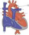
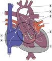
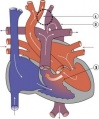
Abnormal Heart Gallery
- HeartILP draft aorticstenosis.jpg
Aortic Stenosis
- HeartILP draft asd.jpg
ASD
Coarctation of the Aorta
- HeartILP draft dorv.jpg
DORV
Functional Hypoplastic Left Heart
- HeartILP draft hlh.jpg
Hypoplastic Left Heart
Interrupted Aortic Arch
- HeartILP draft papvd.jpg
PAPVD
- HeartILP draft patentda.jpg
Patent Ductus Arteriosus
- HeartILP draft pulmonaryatresia.jpg
Pulmonary Atresia
- HeartILP draft pulmonarystenosis.jpg
Pulmonary Stenosis
- HeartILP draft tapvc.jpg
TAPVC
- HeartILP draft tetralogyoffallot.jpg
Tetralogy of Fallot
- HeartILP draft transposition.jpg
Transposition of the Great Vessels
- HeartILP draft tricuspidatresia.jpg
Tricuspid Atresia
- HeartILP draft vsd.jpg
VSD


