Uploads by Z5229431
From Embryology
This special page shows all uploaded files.
| Date | Name | Thumbnail | Size | Description | Versions |
|---|---|---|---|---|---|
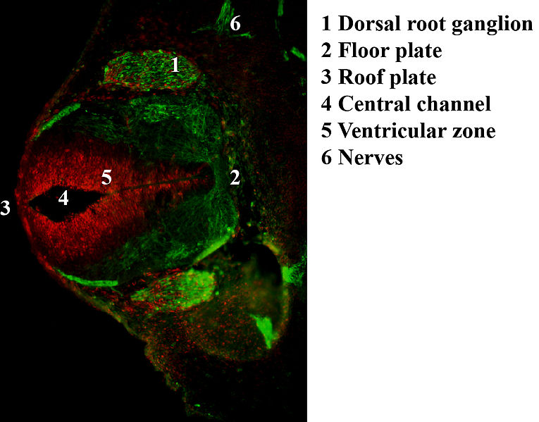
| 18:50, 16 October 2018 | Embryonic dorsal root ganglia in mouse.jpg (file) |  |
84 KB | Antibody stain against Neurofilament (green) and Ki 67 (red) in a Mouse embryo at day 12.5 after fertilization. Shown is the dorsal root ganglion (green ellipsoid regions where cells express neurofilament) and the ventricular zone (red region where cel... | 1 |
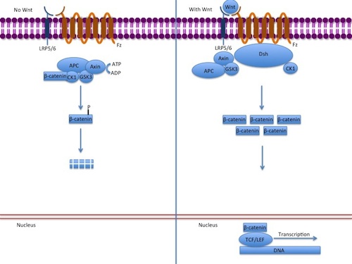
| 17:45, 16 October 2018 | Canonical Wnt pathway.jpg (file) |  |
42 KB | Diagram of the canonical Wnt pathway in the presence and absence of Wnt | 1 |
| 17:39, 15 October 2018 | Illustration of Sox signalling pathway.jpg (file) |  |
50 KB | The diagram illustrates the signalling pathway taken by the transcription factor Sox10 and its partner factor Pouf3f1/2, which activates the Erg2 gene that serves as the second partner factor to express myelin genes during Schwann cell development. | 1 |
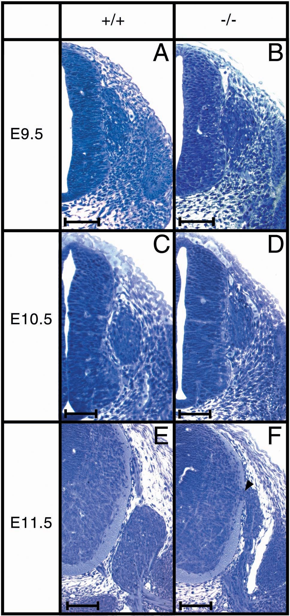
| 14:49, 15 October 2018 | Structure of DRG in Sox10-deficient mice.jpg (file) |  |
441 KB | Histology of DRG in Sox10-deficient mice. Transverse sections, 6 μm through the thoracic neural tube and DRG from 9.5 to 11.5 dpc embryos were stained with toluduine blue. Wildtype (+/+ in A, C, E) and homozygous Sox10-deficient embryos (−/− in B,... | 1 |
| 16:23, 10 September 2018 | Neural crest migration and somite development in zebrafish.jpeg (file) |  |
119 KB | Myotome is the major component of zebrafish somites. Slow muscle precursors (red), called adaxial cells, are located adjacent to notochord before segmentation (left). During segmentation stages they become a single-layered sheet of fully extended muscl... | 1 |
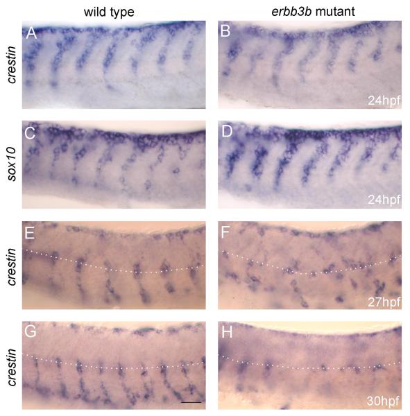
| 01:28, 28 August 2018 | Neural crest cell migration in erbb3b mutants.jpg (file) |  |
45 KB | Neural crest cell migration is initially normal but later disrupted in erbb3b mutants Whole-mount labeling using crestin (A,B,E-H) or sox10 (C,D) riboprobes of wild-type embryos (A,C,E,G) and erbb3b mutants (B,D,F,H). At 24 hpf, both probes show that... | 1 |
| 11:56, 7 August 2018 | Neuropore cell shape change 99.png (file) |  |
1.01 MB | Fig. 3 Cell shape changes in the midline after mid-hindbrain neuropore (MHNP) closure in vivo. a The relationship between the closing of MHNP and the somite stage in ICR strain mouse. b-f Dorsal views of embryos stained with E-cadherin (Ecad). Non-neur... | 1 |