Uploads by Z5229132
From Embryology
This special page shows all uploaded files.
| Date | Name | Thumbnail | Size | Description | Versions |
|---|---|---|---|---|---|
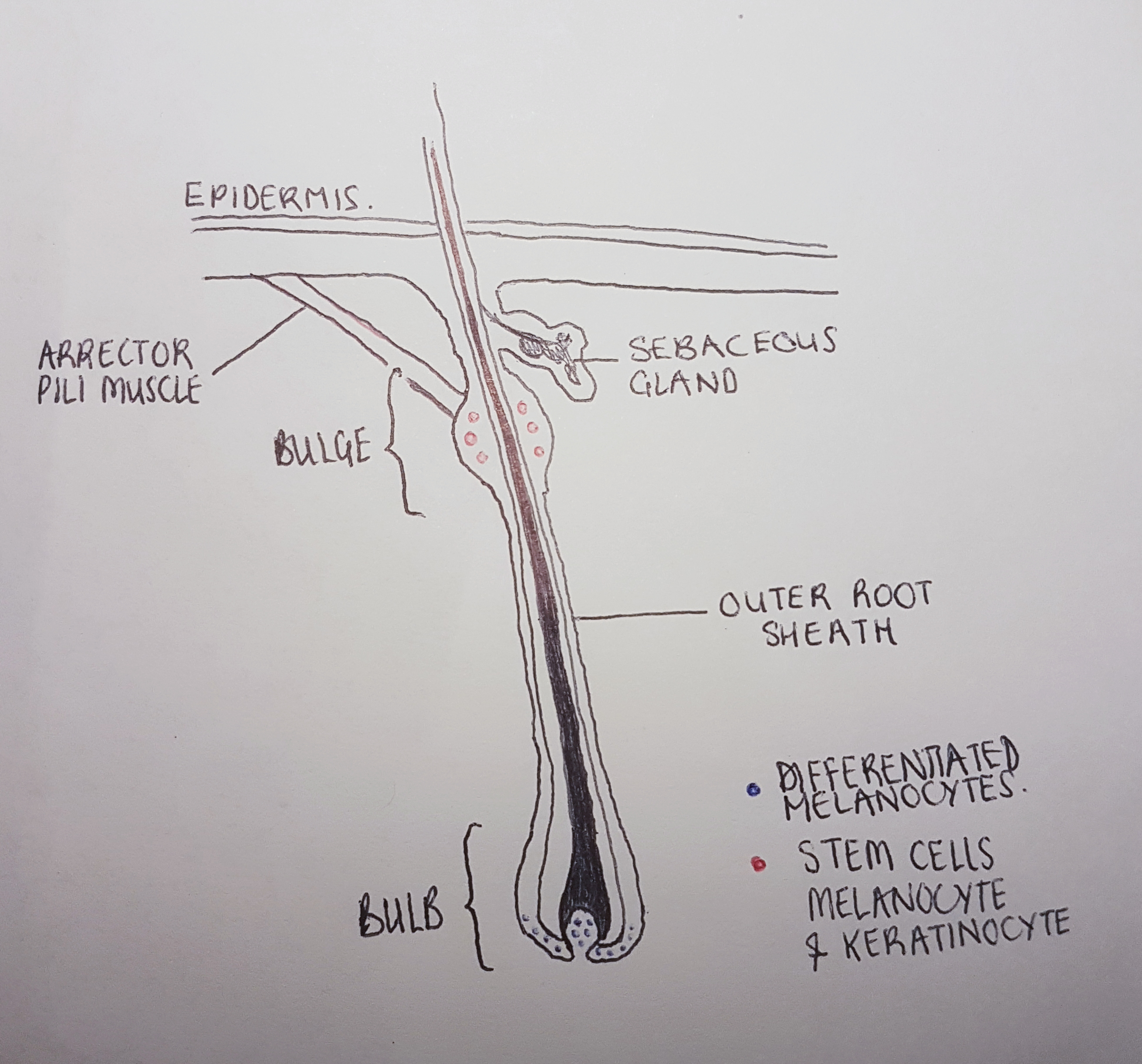
| 21:48, 16 October 2018 | Hair Follicle.jpg (file) |  |
2.14 MB | Diagram of the hair follicle at anagen phase: populations of melanocyte and keratinocyte stem cells are located in the hair bulge in red. Differentiated melanocytes in the hair bulb are in blue. These supply melanosomes for hair pigmentation to the ke... | 1 |
| 20:21, 9 October 2018 | Skin Melanocytes.jpg (file) |  |
79 KB | Diagram of the skin epidermis: shows a melanocyte in the basal epithelium. Its dendrites extend into the stratum spinosum, transferring melanosomes to surrounding keratinocytes via the extracellular space. Melanosomes populate the perinuclear space of... | 2 |
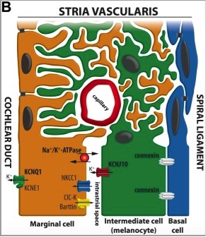
| 17:57, 4 September 2018 | Stria Vascularis diagram 2.jpeg (file) |  |
46 KB | Diagram of the Stria Vascularis, containing the marginal cells, intermediate melanocytes and basal cells. It shows marginal cell extensions intercalating with melanocytes, and the K+ ion channels involved in generating the EP. ====Copyright==== Copyri... | 1 |
| 09:00, 28 August 2018 | Left and Right Cochlea.jpeg (file) |  |
3.45 MB | The left and right cochlea of viable dominant spotting mutant mice, showing asymmetrical pigmentation of ears. The left cochlea has melanocytes in the Stria Vascularis (shown as dots in the centre of the stria) whilst the right cochlea has none. Crosse... | 1 |
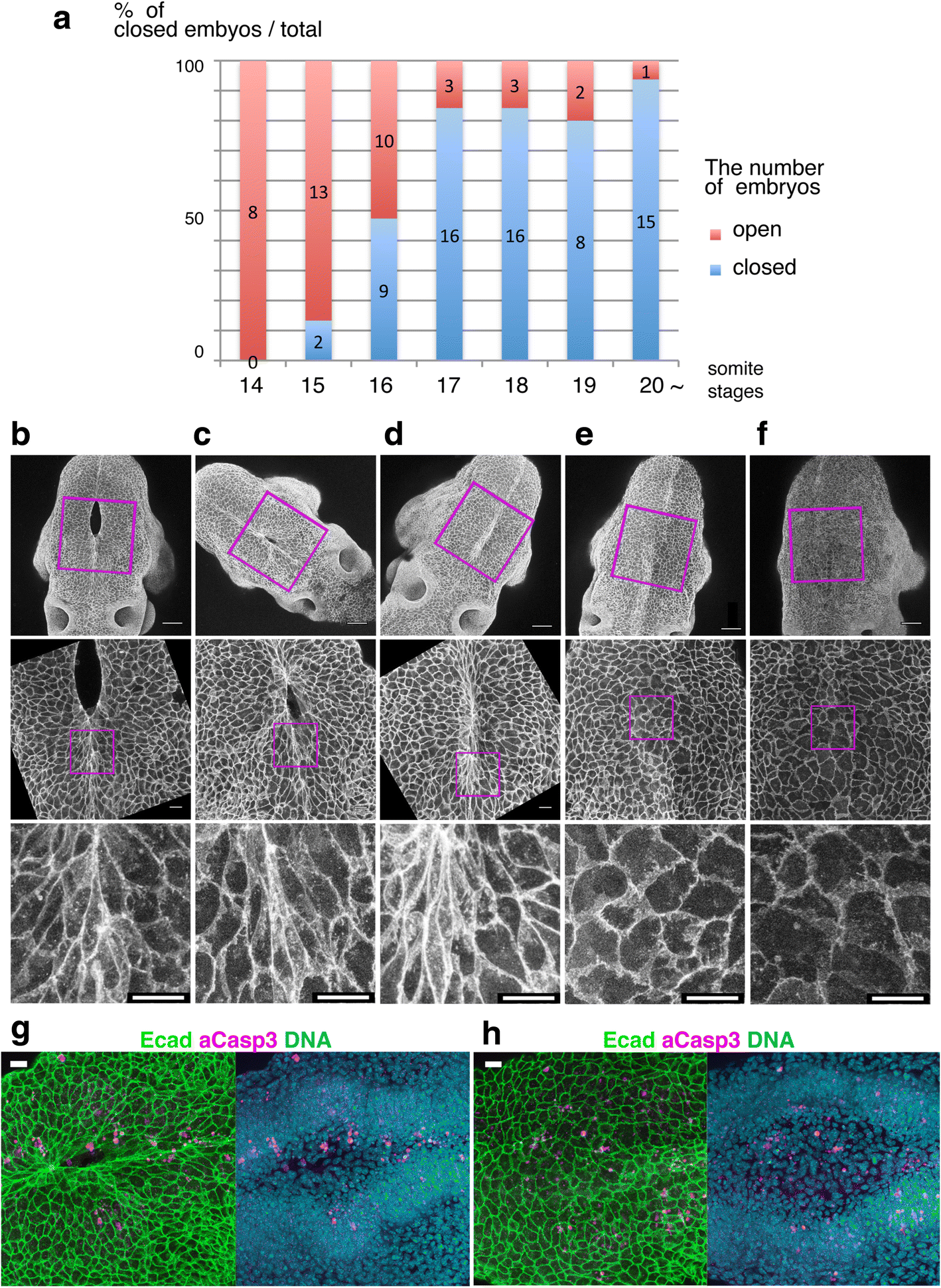
| 11:49, 7 August 2018 | Neuralpore.png (file) |  |
1.01 MB | Cell shape changes in the midline after mid-hindbrain neuropore (MHNP) closure in vivo. a The relationship between the closing of MHNP and the somite stage in ICR strain mouse. b-f Dorsal views of embryos stained with E-cadherin (Ecad). Non-neural ecto... | 1 |