Uploads by Z5164785
From Embryology
This special page shows all uploaded files.
| Date | Name | Thumbnail | Size | Description | Versions |
|---|---|---|---|---|---|
| 10:56, 17 October 2018 | Wnt signalling.jpg (file) |  |
94 KB | Figure 11: Wnt signalling in melanocyte differentiation Confocal microscopy image illustrating how Wnt signalling remains active throughout the process of melanocyte differentiation. ====Reference==== Vibert L, Aquino G, Gehring I, Subkankulova T,... | 2 |
| 10:53, 17 October 2018 | Wnt confocal microscopy.png (file) |  |
200 KB | Figure 11: Wnt signalling in melanocyte differentiation Confocal microscopy image illustrating how Wnt signalling remains active throughout the process of melanocyte differentiation. ====Reference==== Vibert L, Aquino G, Gehring I, Subkankulova T,... | 1 |
| 09:04, 17 October 2018 | Common Vitiligo.jpg (file) |  |
87 KB | Figure 18: Common Vitiligo The image shows the depigmented regions on the skin of a patient with common vitiligo. The lesions can be seen throughout the upper body in varying degrees of size and depigmentation. ====Reference==== Faria AR, Tarlé RG,... | 1 |
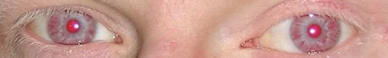
| 23:09, 16 October 2018 | Oculocutaneous albinism eyes.jpg (file) | 15 KB | Figure 20: OCA1A patient eyes The image shows the eyes of a patient with OCA1A. The iris is observed to be fully translucent and an ‘almost pink’ colour due to the lack of pigment development. ====Reference==== Grønskov, K., Ek, J. and Brondum-N... | 1 | |
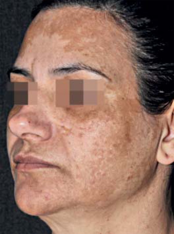
| 22:45, 16 October 2018 | Mixed melasma.jpg (file) |  |
112 KB | Figure 17: Mixed Facial Melasma The image shows the mixed facial melasma of a female patient. The melasma is described as mixed as it is a mixture of the identified facial patterns, affecting the frontal, temporal, parotid, mandibular and zygomatic re... | 2 |
| 21:55, 16 October 2018 | Leopard syndrome skin appearance.jpeg (file) |  |
88 KB | Figure 12: The appearance of 4 different Leopard syndrome affected individuals, all of different ages. ===Reference=== {{#pmid:18505544|PMID18505544}} ====Copyright==== © 2008, Sarkozy et al; licensee BioMed Central Ltd. {{Student Image}} | 1 |
| 21:51, 16 October 2018 | Leopard syndrome skin appearance.jpg (file) |  |
88 KB | Figure 12: The skin appearance of 4 different Leopard syndrome affected individuals, all of different ages. Lentigines seen at different stages. ===Reference=== {{#pmid:18505544|PMID18505544}} ====Copyright==== © 2008, Sarkozy et al; licensee BioMe... | 1 |
| 21:42, 16 October 2018 | Leopard syndrome lentigines.jpg (file) |  |
88 KB | Lentigine appearance on the skin of LS patients at different ages | 1 |
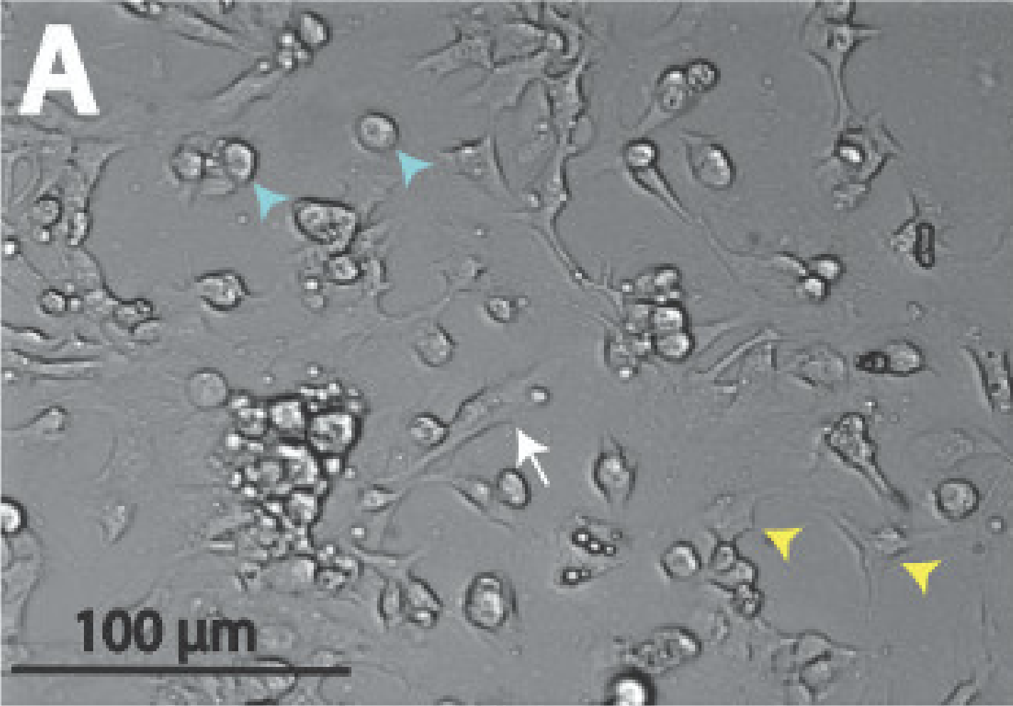
| 23:10, 28 August 2018 | Cardiac melanocyte in an embryonic mouse pup.png (file) |  |
427 KB | 400px Figure 3. A differential interference contrast (DIC) microscopical image of a cardiac melanocyte present in an isolated atrial cell of an embryonic mouse pup. The white arrow indicates... | 1 |