Uploads by Z3332824
From Embryology
This special page shows all uploaded files.
| Date | Name | Thumbnail | Size | Description | Versions |
|---|---|---|---|---|---|
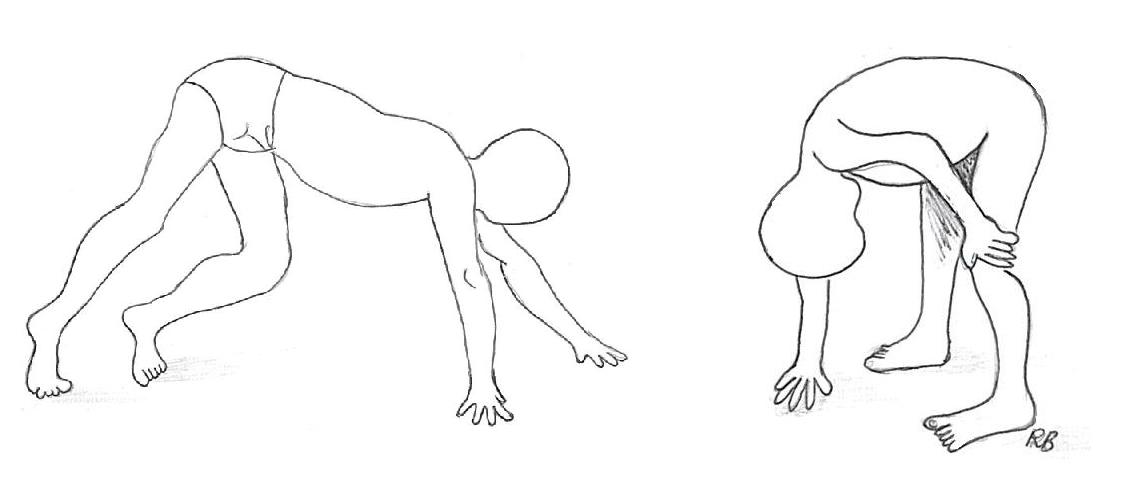
| 14:53, 7 October 2011 | Gower's sign - a symptom of DMD.JPG (file) |  |
36 KB | Gower's sign - a symptom of Duchenne Muscular Dystrophy (DMD). This picture is of a boy afflicted with DMD - he struggles to stand due to his weak skeletal muscles. The second boy is using his arms to push himself up along his legs in an attempt to stand | 2 |
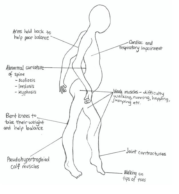
| 11:46, 6 October 2011 | Symptoms of DMD.JPG (file) |  |
39 KB | Symptoms exhibited in sufferers of Duchenne Muscular Dystrophy (DMD). This picture is a drawing of a boy suffering from DMD and the typical symptoms associated with this congenital disease. Created by z3332824 for use on the group research assignment of | 1 |
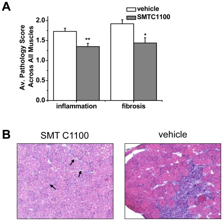
| 13:39, 2 October 2011 | Utrophin effects compared to control.jpg (file) |  |
114 KB | Effects of utrophin upregulation on muscle cells, inflammation and fibrosis in comparison to the control (vehicle group). (A) Data demonstrates the reduction in overall skeletal muscle inflammation and fibrosis from mdx treated with SMT C1100 compared t | 1 |
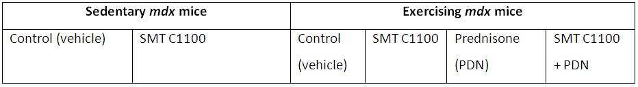
| 21:02, 1 October 2011 | Experiment groups for utrophin upregulation.JPG (file) | 20 KB | Groups used in utrophin upregulation experiment. This table summarises the experiment groups used by Tinsley & Fairclough et. al (2011) in their experiments on mdx mice. Two main groups of mice were organised - one was exercising, the other was sedent | 2 | |
| 20:33, 1 October 2011 | Tinsley et al. utrophin results.jpg (file) |  |
114 KB | Reduction in pathological features of DMD from use of utrophin up-regulation. Reduction in secondary pathological features. (A) Data demonstrates the reduction in overall skeletal muscle inflammation and fibrosis from mdx treated with SMT C1100 compared | 1 |
| 22:18, 16 September 2011 | Spinal problems DMD.jpg (file) |  |
127 KB | A 11-year-old boy, wheelchair bound for 8 months with Duchenne muscular dystrophy. (A) Preoperative antero-posterior radiograph right sided showing 60° curve. (B, C) Two year post-operative antero-posterior and lateral radiographs showing sublaminar wiri | 1 |
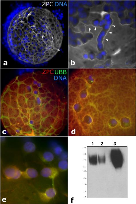
| 10:54, 25 August 2011 | Patterns of zona pellucida deposition.JPG (file) |  |
44 KB | ==Patterns of zona pellucida deposition== (a, b) Accumulation of ZPC (gray) in ridges (arrowheads) adjacent to zona-adhering corona radiata cells (blue = nuclei stained with DAPI). (c-e) Colocalization of ZPC (red) and ubiquitin (green) in the zona pellu | 1 |