Uploads by Z3292953
From Embryology
This special page shows all uploaded files.
| Date | Name | Thumbnail | Size | Description | Versions |
|---|---|---|---|---|---|
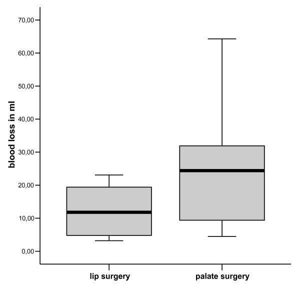
| 10:54, 11 October 2011 | Blood Loss During Lip and Palate Repair.jpg (file) |  |
21 KB | Figure 1 Directly measured blood loss during primary lip(n = 28) and palate repair (n = 40). This is an Open Access article distributed under the terms of the Creative Commons Attribution License (http://creativecommons.org/licenses/by/2.0), which permit | 1 |
| 10:42, 11 October 2011 | Cleft Palate Maxillary and Mandibular View.jpg (file) |  |
141 KB | Figure 1 Cleft palate - maxillary and mandibular view This is an open-access article distributed under the terms of the Creative Commons Attribution-Noncommercial-Share Alike 3.0 Unported, which permits unrestricted use, distribution, and reproduction in | 1 |
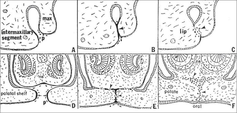
| 10:36, 11 October 2011 | Comparison of morphogenesis of the upper lip with the palate.jpg (file) |  |
188 KB | Figure 2 Comparison of the morphogenesis of the upper lip (A-C) with that of the palate (D-F). After the bilateral maxillary processes (max) fuse externally with the inter-maxillary segment, the resulting epithelial seam (arrow, B) gives rise to mesenchym | 1 |
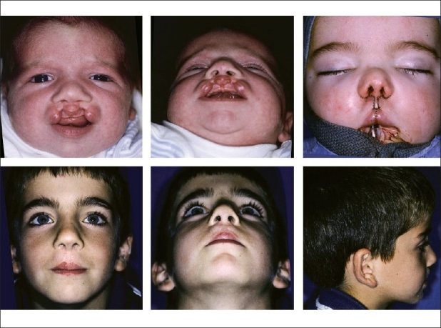
| 10:24, 11 October 2011 | Bilateral Cleft Lip With Nasal Deformity.jpg (file) |  |
148 KB | Figure 1 Findings in bilateral cleft lip nasal deformity This is an open-access article distributed under the terms of the Creative Commons Attribution License, which permits unrestricted use, distribution, and reproduction in any medium, provided the or | 1 |
| 10:13, 11 October 2011 | From infancy until completion of treatment.jpg (file) |  |
103 KB | Figure 8B (B) Right lateral view from infancy until completion of treatment This is an open-access article distributed under the terms of the Creative Commons Attribution License, which permits unrestricted use, distribution, and reproduction in any medi | 1 |
| 13:03, 6 October 2011 | Repair of Bilateral Cleft Lip.jpg (file) |  |
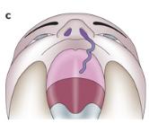
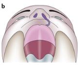
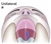
179 KB | Figure 9 (A). Infant with bilateral symmetrical incomplete cleft lip and intact secondary palate, (B) Submental view, (C) Following synchronous nasolabial repair, including positioning and fixation lower lateral cartilages, (A, B & C) Appearance at age si | 1 |
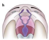
| 18:05, 29 September 2011 | Bilateral cleft lip with cleft hard and soft palate.jpg (file) |  |
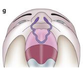
5 KB | Figure 2 Types of cleft A:A set of illustrative drawings of CLP types.f, g and h show degrees of bilateral cleft lip and palate. <pubmed>21331089</pubmed> Permission: Adapted by permission from Macmillan Publishers Ltd: [Nature] (Michael J. Dixon,1 Mary | 1 |
| 18:04, 29 September 2011 | Bilateral cleft lip with cleft hard palate.jpg (file) |  |
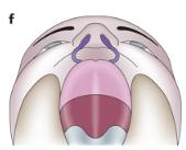
5 KB | Figure 2 Types of cleft A:A set of illustrative drawings of CLP types.f, g and h show degrees of bilateral cleft lip and palate. <pubmed>21331089</pubmed> Permission: Adapted by permission from Macmillan Publishers Ltd: [Nature] (Michael J. Dixon,1 Mary | 1 |
| 18:02, 29 September 2011 | Bilateral cleft lip.jpg (file) |  |
5 KB | Figure 2 Types of cleft A:A set of illustrative drawings of CLP types.f, g and h show degrees of bilateral cleft lip and palate. <pubmed>21331089</pubmed> Permission: Adapted by permission from Macmillan Publishers Ltd: [Nature] (Michael J. Dixon,1 Mary | 1 |
| 17:59, 29 September 2011 | Type Bilateral cleft palate.jpg (file) |  |
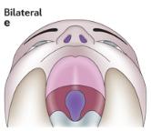
5 KB | Figure 2 Types of cleft A: A set of illustrative drawings of CLP types.111 a and e show unilateral and bilateral clefts of the soft palate <pubmed>21331089</pubmed> Permission: Adapted by permission from Macmillan Publishers Ltd: [Nature] (Michael J. Di | 1 |
| 17:43, 29 September 2011 | Unilateral cleft lip with cleft hard and soft palate.jpg (file) |  |
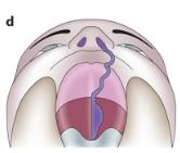
5 KB | Figure 2 Types of cleft A: A collection of images of different types of clefts, some with associated anomalies such as lip pits.111 a–c <pubmed>21331089</pubmed> Permission: Adapted by permission from Macmillan Publishers Ltd: [Nature] (Michael J. Dix | 1 |
| 17:40, 29 September 2011 | Unilateral cleft lip with a cleft hard palate.jpg (file) |  |
5 KB | Figure 2 Types of cleft A: A collection of images of different types of clefts, some with associated anomalies such as lip pits.111 a–c <pubmed>21331089</pubmed> Permission: Adapted by permission from Macmillan Publishers Ltd: [Nature] (Michael J. Dix | 1 |
| 17:39, 29 September 2011 | Unilateral cleft lip.jpg (file) |  |
5 KB | Figure 2 Types of cleft A: A collection of images of different types of clefts, some with associated anomalies such as lip pits.111 a–c <pubmed>21331089</pubmed> Permission: Adapted by permission from Macmillan Publishers Ltd: [Nature] (Michael J. Dix | 1 |
| 17:33, 29 September 2011 | Type Unilateral Cleft Palate.jpg (file) |  |
5 KB | Figure 2 Types of cleft A: A collection of images of different types of clefts, some with associated anomalies such as lip pits.111 a–c <pubmed>21331089</pubmed> Permission: Adapted by permission from Macmillan Publishers Ltd: [Nature] (Michael J. Dix | 1 |
| 16:05, 14 September 2011 | Normal palate shelf and key stages of mouse palatal development.jpg (file) | 80 KB | Figure 1 Schematic drawing showing coronal view of a normal palate shelf and key stages of mouse palatal development. At E12-E13 days in the mouse gestation, the palatal shelves grow downward along the tongue (t). At E13-E13.5 days, the palatal shelves be | 1 | |
| 13:05, 12 September 2011 | Bilateral Cleft Lip Variations.jpg (file) |  |
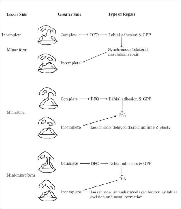
87 KB | Figure 10 Algorithm for repair of bilateral asymmetrical cleft lip (complete or incomplete) and contralateral incomplete or lesser-form cleft (Modified from Yuzuriha, Oh, Mulliken, 2008) John B. Mulliken,Repair of bilateral cleft lip and its variants,Ind | 1 |
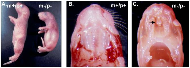
| 12:00, 16 August 2011 | File-Cleft palate in newborn mice.jpg (file) |  |
26 KB | A. Growth retardation of 2-day-old pup with a homozygous deletion from Ube3a to Gabrb3. B & C. Cleft palate in a pup with a homozygous deletion from Ube3a to Gabrb3. The black arrow points to the cleft palate in pup with a homozygous deletion (m−/p−). | 1 |