Uploads by Z3284061
From Embryology
This special page shows all uploaded files.
| Date | Name | Thumbnail | Size | Description | Versions |
|---|---|---|---|---|---|
| 00:15, 13 October 2011 | Pie Chart.JPG (file) |  |
50 KB | This Pie Chart is created by Student z3308965 Pie chart shows the mortality rate in different countries. {{Template:2011 Student Image}} | 1 |
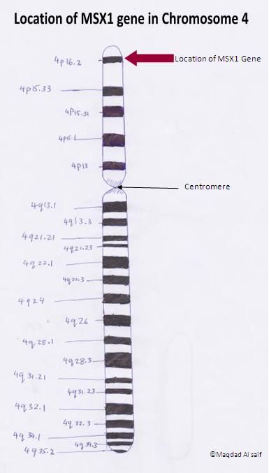
| 14:23, 12 October 2011 | MSX 1 Gene.JPG (file) |  |
22 KB | The Image is drawn by student z3284061 Image shows the Location of MSX1 gene in Chromosome 4. The Image is based on information in the article. '''Reference''' <pubmed>9369446</pubmed> {{Template:2011 Student Image}} | 1 |
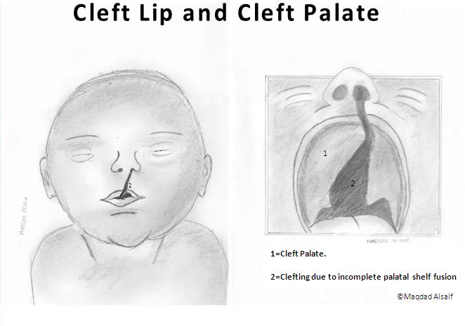
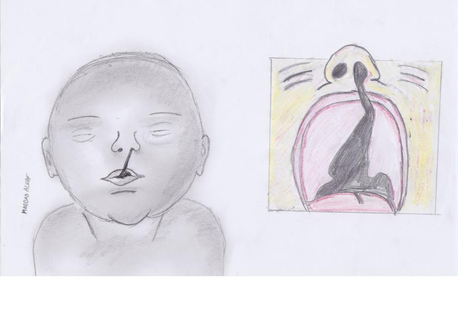
| 13:05, 12 October 2011 | Oral Clefting.JPG (file) |  |
31 KB | This image was drawn by student z3284061. The image shows an example of Oral Clefting. {{Template:2011 Student Image}} | 1 |
| 20:59, 10 October 2011 | Modified prominences final.jpg (file) |  |
23 KB | This image is drawn by Student Maqdad Alsaif ,z3284061. Diagram Shows 5-week embryo with five major prominences. {{Template:2011 Student Image}} | 1 |
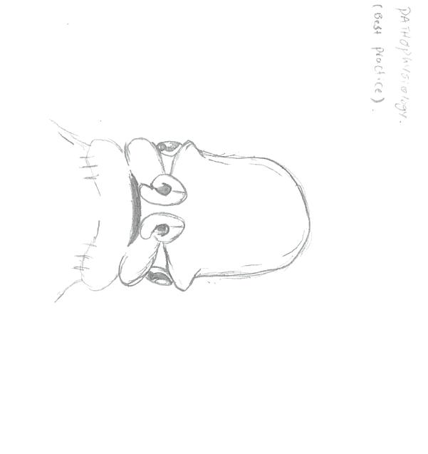
| 14:59, 9 October 2011 | Pathophysiology & development.jpg (file) |  |
15 KB | Image drawn by student z3284061. Diagram of the 5-week old embryo with the 5 major facial prominences. | 1 |
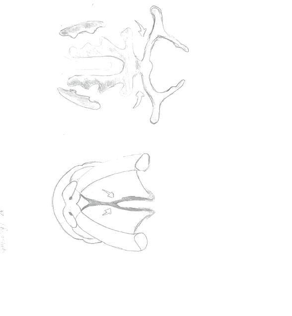
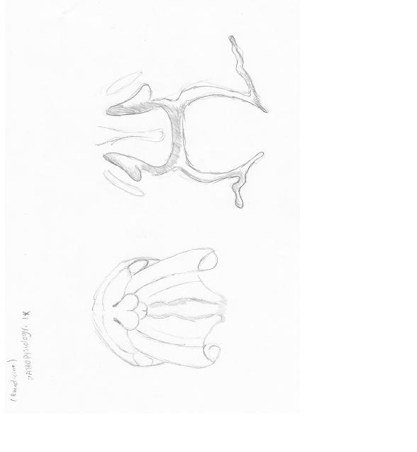
| 14:56, 9 October 2011 | Pathophysiology 2.jpg (file) |  |
17 KB | Image drawn by student z3284061 Palatal shelves grow downward and adjacent to the tongue. | 1 |
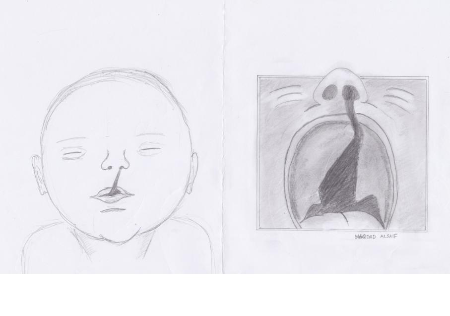
| 14:54, 9 October 2011 | Pathophyis 1.jpg (file) |  |
16 KB | image drawn by z3284061 Description: Embryology processes of abnormal palatal development. Normally, the palatal shelves assume a vertical orientation and are directed downward. Free communication exists between the oral cavity and the nose at 7 weeks. T | 1 |
| 21:44, 8 October 2011 | Figure 3. Fetal Lip and Primary Palate Three dimensional versus Two-dimensional US.gif (file) |  |
126 KB | Figure 3. Three-dimensional rendered frontal oblique US image shows a median cleft lip (arrow) in a fetus at 32 weeks gestational age. Dear Maqdad Alsaif: The Radiological Society of North America (RSNA®) is pleased to grant you permission to reproduc | 2 |
| 21:40, 8 October 2011 | Figure 1. Fetal Lip and Primary Palate Three dimensional versus Two dimensional US.gif (file) |  |
129 KB | Figure 1. Three-dimensional rendered US image viewed frontally shows a facial cleft (arrows) in a fetus at 22 weeks gestational age. After viewing the rotating 3D image, the family elected to continue the pregnancy.<ref name="PMID11012450"><pubmed>110124 | 2 |
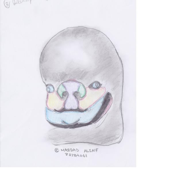
| 20:48, 8 October 2011 | Development 3.jpg (file) |  |
21 KB | This Image is drawn by the student Maqdad Alsaif- z3284061 {{Template:2011 Student Image}} | 1 |
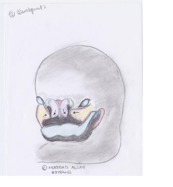
| 20:47, 8 October 2011 | Development 2.jpg (file) |  |
22 KB | This Image is drawn by the student Maqdad Alsaif- z3284061 {{Template:2011 Student Image}} | 1 |
| 20:47, 8 October 2011 | Development1.jpg (file) |  |
22 KB | This Image is drawn by the student Maqdad Alsaif- z3284061 {{Template:2011 Student Image}} | 1 |
| 20:41, 8 October 2011 | Introduction 4.jpg (file) |  |
31 KB | This Image is drawn by the student Maqdad Alsaif- z3284061 {{Template:2011 Student Image}} | 1 |
| 20:41, 8 October 2011 | Introduction 3.jpg (file) |  |
40 KB | This Image is drawn by the student Maqdad Alsaif- z3284061 {{Template:2011 Student Image}} | 1 |
| 20:19, 8 October 2011 | Figure 2. Fetal Lip and Primary Palate Three dimensional versus Two dimensional US.gif (file) |  |
120 KB | Figure 2. A, Coronal, B, sagittal, C, transverse, and D, 3D rendered US images of a cleft lip (arrows) and palate in a fetus at 21 weeks gestational age. Image in D is viewed frontally. White line indicates plane depicted in C. Dear Maqdad Alsaif: The | 1 |
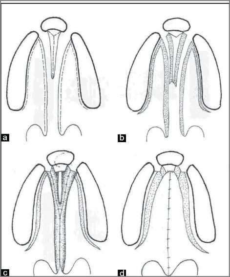
| 15:44, 7 October 2011 | Line diagram showing Bardach two-flap technique of palatoplasty in a bilateral cleft lip and palate.jpg (file) |  |
50 KB | Line diagram showing Bardach two-flap technique of palatoplasty in a bilateral cleft lip and palate. This is a modification of the von Langenbeck technique in which the incision is made along the cleft margin and the alveolar margin. These are joined ante | 7 |
| 10:50, 15 September 2011 | Variations of Cleft Lip or Palate.jpg (file) |  |
445 KB | Figure 2 Types of cleft A: A collection of images of different types of clefts, some with associated anomalies such as lip pits.111 a–c, Van der Woude syndrome cases with associated lip pits; d, isolated cleft palate only; e, isolated unilateral cleft l | 1 |
| 17:25, 9 September 2011 | Cleft lip 2.jpg (file) |  |
160 KB | This Image shows bilateral cleft lip Before Surgery. Source: http://www.flickr.com/photos/interplast/63952577/ Creative Commons- Attribution-NonCommercial-NoDerivs 2.0 Generic (CC BY-NC-ND 2.0) You are free: to Share — to copy, distribute and tr | 1 |
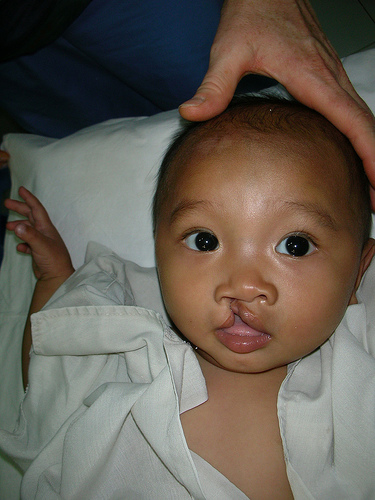
| 17:17, 9 September 2011 | Cleft lip.jpg (file) |  |
101 KB | This image shows Pre-Op Unilateral Cleft Lip. Source: http://www.flickr.com/photos/interplast/131093645/ Creative Commons- Attribution-NonCommercial-NoDerivs 2.0 Generic (CC BY-NC-ND 2.0) You are free: to Share — to copy, distribute and transmit | 2 |
| 13:01, 11 August 2011 | 468px-Differentially expressed RefSeq genes in human trisomy 21.jpg (file) |  |
61 KB | Pone.0018493.g006.jpg http://www.ncbi.nlm.nih.gov/pmc/articles/PMC3080369/ Figure 6 Differentially expressed RefSeq genes in human trisomy 21. (A) Standard MA-plot of the normalized global observed counts per each RefSeq gene. (B) shows the percentage o | 2 |