Uploads by Z3217015
From Embryology
This special page shows all uploaded files.
| Date | Name | Thumbnail | Size | Description | Versions |
|---|---|---|---|---|---|
| 12:39, 24 September 2009 | Drosophila table.JPG (file) |  |
52 KB | Drosophila development table. Image courtesy of Katrin Weigmann, Robert Klapper, Thomas Strasser, Christof Rickert, Gerd Technau, Herbert Jäckle, Wilfried Janning and Christian Klämbt: FlyMove – a new way to look at development of Drosophila.Trends G | 1 |
| 12:32, 24 September 2009 | Drosophila Musculature.JPG (file) |  |
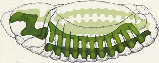
26 KB | The muscle system in the stage 13 embryo. Dorso-Lateral aspect. Oriented anterior left. After Somatic Musculature 13 dorsolateral view on page 38 from Hartenstein (1993). V. Hartenstein. Atlas of Drosophila development. Cold Spring Harbor Laboratory Press | 1 |
| 12:52, 18 September 2009 | Stage 8 drosophila .gif (file) |  |
18 KB | Stage 8 Drosophila embryo. Scanning electron microscope image of dorsal view. Turner, F.R. and Mahowald, A.P. (1979). Scanning electron microscopy of Drosophila melanogaster embryogenesis. III. Formation of the head and caudal segments. Dev. Biol. 68: 96 | 1 |
| 12:46, 18 September 2009 | Stage 17 drosophila.jpg (file) |  |
29 KB | Stage 17 Drosophila embryo. The section is stained using an anti-Crumbs antibody, showing epithelial structures. Image courtesy of Katrin Weigmann, Robert Klapper, Thomas Strasser, Christof Rickert, Gerd Technau, Herbert Jäckle, Wilfried Janning and Chr | 1 |
| 12:44, 18 September 2009 | Stage 15 drosophila.jpg (file) |  |
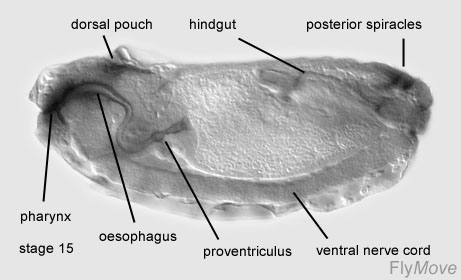
26 KB | Stage 15 Drosophila embryo. The section is stained using an anti-Crumbs antibody, showing epithelial structures. Image courtesy of Katrin Weigmann, Robert Klapper, Thomas Strasser, Christof Rickert, Gerd Technau, Herbert Jäckle, Wilfried Janning and Chr | 1 |
| 12:43, 18 September 2009 | Stage 14 drosophila.jpg (file) |  |
22 KB | Stage 14 Drosophila embryo. The section is stained using an anti-Crumbs antibody, showing epithelial structures. Image courtesy of Katrin Weigmann, Robert Klapper, Thomas Strasser, Christof Rickert, Gerd Technau, Herbert Jäckle, Wilfried Janning and Chr | 1 |
| 12:40, 18 September 2009 | Stage 13 drosophila.jpg (file) |  |
24 KB | Stage 13 Drosophila embryo. The section is stained using an anti-Crumbs antibody, showing epithelial structures. Image courtesy of Katrin Weigmann, Robert Klapper, Thomas Strasser, Christof Rickert, Gerd Technau, Herbert Jäckle, Wilfried Janning and Chr | 1 |
| 12:38, 18 September 2009 | Stage 12 drosophila.jpg (file) |  |
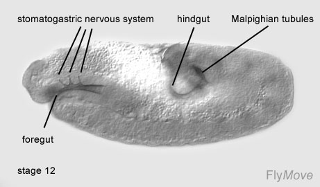
22 KB | Stage 12 Drosophila embryo. The section is stained using an anti-Crumbs antibody, showing epithelial structures. Image courtesy of Katrin Weigmann, Robert Klapper, Thomas Strasser, Christof Rickert, Gerd Technau, Herbert Jäckle, Wilfried Janning and Chr | 1 |
| 12:37, 18 September 2009 | Stgae 11 drosophila.jpg (file) |  |
27 KB | Stage 11 Drosophila embryo. The section is stained using an anti-Crumbs antibody, showing epithelial structures. Image courtesy of Katrin Weigmann, Robert Klapper, Thomas Strasser, Christof Rickert, Gerd Technau, Herbert Jäckle, Wilfried Janning and Chr | 1 |
| 12:36, 18 September 2009 | Stage 10 drosophila.jpg (file) |  |
38 KB | Stage 10 Drosophila embryo. The section is stained using an anti-Crumbs antibody, showing epithelial structures. Image courtesy of Katrin Weigmann, Robert Klapper, Thomas Strasser, Christof Rickert, Gerd Technau, Herbert Jäckle, Wilfried Janning and Ch | 1 |
| 12:35, 18 September 2009 | Stage 9 drosophila.jpg (file) |  |
21 KB | Stage 9 Drosophila embryo. The section is stained using an anti-Crumbs antibody, showing epithelial structures. Image courtesy of Katrin Weigmann, Robert Klapper, Thomas Strasser, Christof Rickert, Gerd Technau, Herbert Jäckle, Wilfried Janning and Chri | 1 |
| 12:32, 18 September 2009 | Stage 7 drosophila.jpg (file) |  |
31 KB | Stage 7 Drosophila embryo. Image courtesy of Katrin Weigmann, Robert Klapper, Thomas Strasser, Christof Rickert, Gerd Technau, Herbert Jäckle, Wilfried Janning and Christian Klämbt: FlyMove – a new way to look at development of Drosophila.Trends Gene | 1 |
| 12:30, 18 September 2009 | Stage 6 drosophila.jpg (file) |  |
28 KB | Stage 6 of Drosophila embryo. Image courtesy of Katrin Weigmann, Robert Klapper, Thomas Strasser, Christof Rickert, Gerd Technau, Herbert Jäckle, Wilfried Janning and Christian Klämbt: FlyMove – a new way to look at development of Drosophila.Trends G | 1 |
| 12:27, 18 September 2009 | Drosophila stage 5.jpg (file) |  |
29 KB | Stage 5 Drosophila embryo. Mid-saggital section Image courtesy of Katrin Weigmann, Robert Klapper, Thomas Strasser, Christof Rickert, Gerd Technau, Herbert Jäckle, Wilfried Janning and Christian Klämbt: FlyMove – a new way to look at development of D | 1 |
| 12:26, 18 September 2009 | Drosophila stage 4.jpg (file) |  |
30 KB | Stage 4 of the Drosophila embryo Image courtesy of Katrin Weigmann, Robert Klapper, Thomas Strasser, Christof Rickert, Gerd Technau, Herbert Jäckle, Wilfried Janning and Christian Klämbt: FlyMove – a new way to look at development of Drosophila.Trend | 1 |
| 12:24, 18 September 2009 | Stage 3 drosophila.jpg (file) |  |
26 KB | Stage 3 Drosophila embryo. Image courtesy of Katrin Weigmann, Robert Klapper, Thomas Strasser, Christof Rickert, Gerd Technau, Herbert Jäckle, Wilfried Janning and Christian Klämbt: FlyMove – a new way to look at development of Drosophila.Trends Gene | 1 |
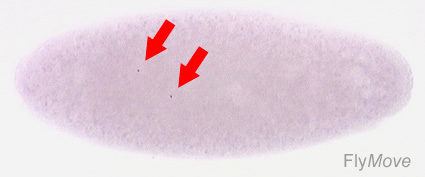
| 12:19, 18 September 2009 | Drosophila stage 2.jpg (file) |  |
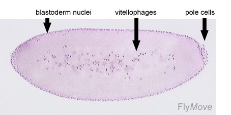
20 KB | The Stage 2 Drosophila Embryo. Peripheral nuclei are visible. Central nuclei (marked with arrows) form vitellophages. Image courtesy of Katrin Weigmann, Robert Klapper, Thomas Strasser, Christof Rickert, Gerd Technau, Herbert Jäckle, Wilfried Janning an | 1 |
| 12:16, 18 September 2009 | Drosophila stage 1.jpg (file) |  |
20 KB | The Stage 1 Drosophila Embryo. The two nuclei are marked with arrows following the first cleavage division. A fuchsin stain was used. Image courtesy of Katrin Weigmann, Robert Klapper, Thomas Strasser, Christof Rickert, Gerd Technau, Herbert Jäckle, Wil | 1 |