File list
From Embryology
This special page shows all uploaded files.
| Date | Name | Thumbnail | Size | User | Description | Versions |
|---|---|---|---|---|---|---|
| 00:51, 25 August 2023 | Wayback machine2004-2023.jpg (file) | 46 KB | Z8600021 | 1 | ||
| 14:36, 23 December 2021 | Australia's mothers and babies 2018.jpg (file) |  |
33 KB | Z8600021 | 1 | |
| 14:23, 23 December 2021 | Assisted reproductive technology in Australia and New Zealand 2019.jpg (file) |  |
68 KB | Z8600021 | 1 | |
| 14:19, 23 December 2021 | Assisted reproductive technology in Australia and New Zealand 2018.jpg (file) |  |
70 KB | Z8600021 | 1 | |
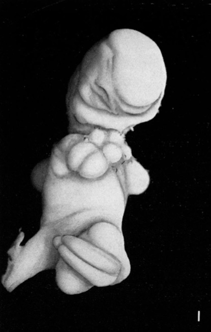
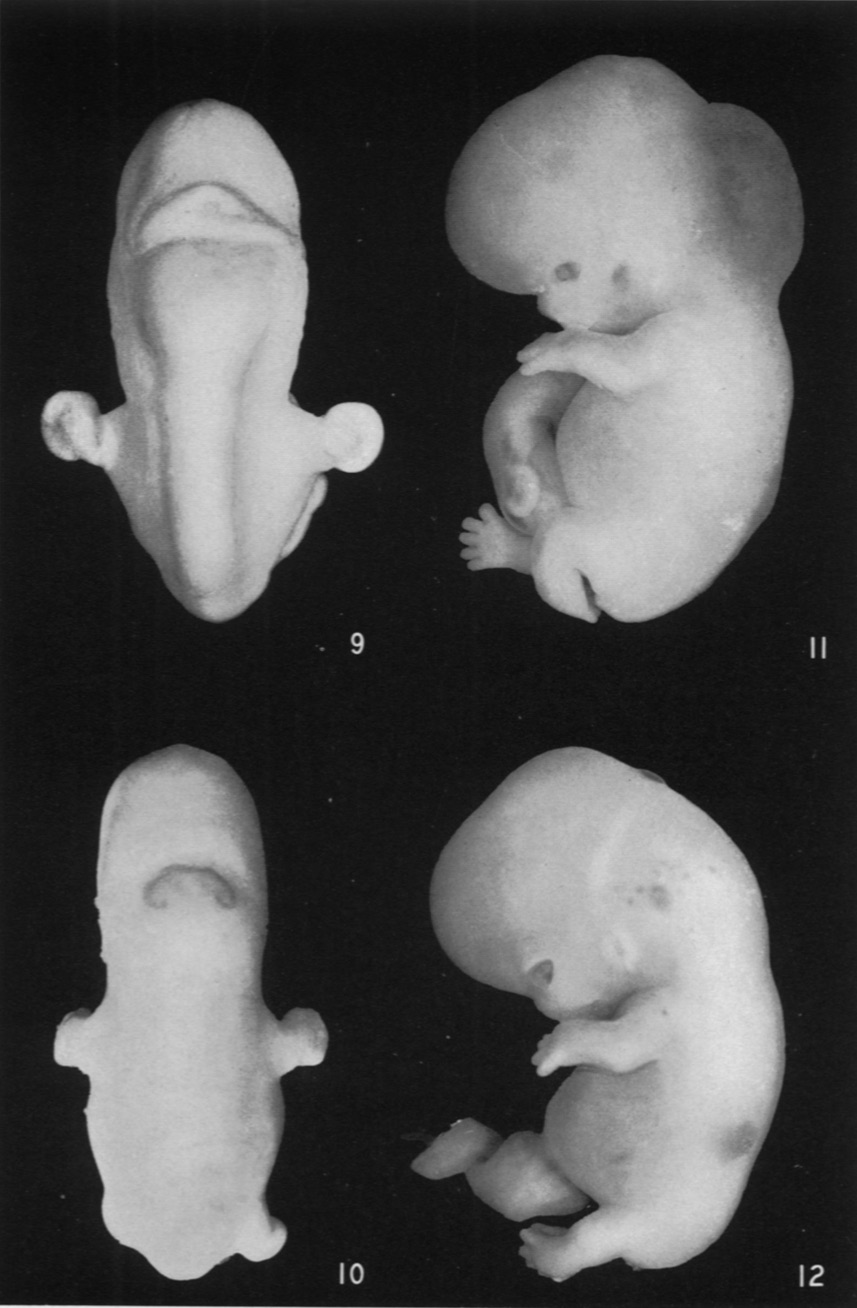
| 09:35, 14 October 2020 | Ingalls1932b fig12.jpg (file) |  |
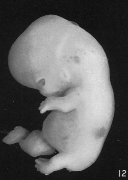
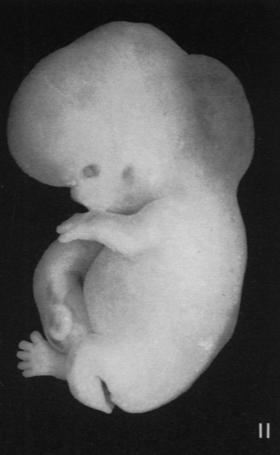
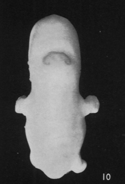
47 KB | Z8600021 | ==Plate 101== Fig. 9. Embryo No. 652. Greatest length 15 mm. Dorsal view of anterior end of embryo to show the transverse discolored band just behind the midbrain. Fig. 10. Embryo No. 167. Greatest length 18.5mm. Very conspicuous, sharply defined, discolored area over the rhombencephalon. There is a similar smaller patch over the vertex and two paired spots on the upper part of the forehead. Fig. 11. Embryo No. 671. Greatest length 25 mm. Enormous blood-stained bleb on back of head and nec... | 1 |
| 09:32, 14 October 2020 | Ingalls1932b fig11.jpg (file) |  |
50 KB | Z8600021 | ==Plate 101== Fig. 9. Embryo No. 652. Greatest length 15 mm. Dorsal view of anterior end of embryo to show the transverse discolored band just behind the midbrain. Fig. 10. Embryo No. 167. Greatest length 18.5mm. Very conspicuous, sharply defined, discolored area over the rhombencephalon. There is a similar smaller patch over the vertex and two paired spots on the upper part of the forehead. Fig. 11. Embryo No. 671. Greatest length 25 mm. Enormous blood-stained bleb on back of head and nec... | 1 |
| 09:32, 14 October 2020 | Ingalls1932b fig10.jpg (file) |  |
41 KB | Z8600021 | ==Plate 101== Fig. 9. Embryo No. 652. Greatest length 15 mm. Dorsal view of anterior end of embryo to show the transverse discolored band just behind the midbrain. Fig. 10. Embryo No. 167. Greatest length 18.5mm. Very conspicuous, sharply defined, discolored area over the rhombencephalon. There is a similar smaller patch over the vertex and two paired spots on the upper part of the forehead. Fig. 11. Embryo No. 671. Greatest length 25 mm. Enormous blood-stained bleb on back of head and nec... | 1 |
| 09:32, 14 October 2020 | Ingalls1932b fig09.jpg (file) |  |
46 KB | Z8600021 | ==Plate 101== Fig. 9. Embryo No. 652. Greatest length 15 mm. Dorsal view of anterior end of embryo to show the transverse discolored band just behind the midbrain. Fig. 10. Embryo No. 167. Greatest length 18.5mm. Very conspicuous, sharply defined, discolored area over the rhombencephalon. There is a similar smaller patch over the vertex and two paired spots on the upper part of the forehead. Fig. 11. Embryo No. 671. Greatest length 25 mm. Enormous blood-stained bleb on back of head and nec... | 1 |
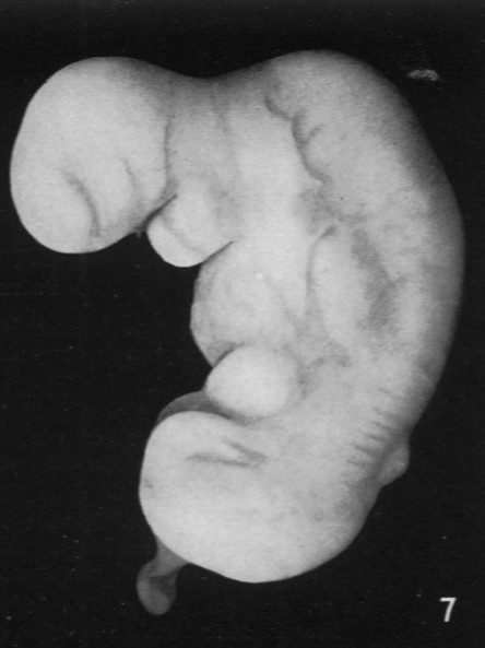
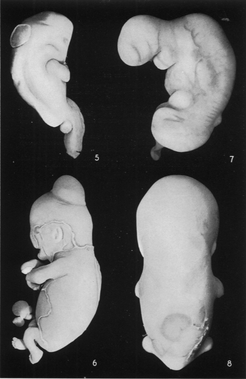
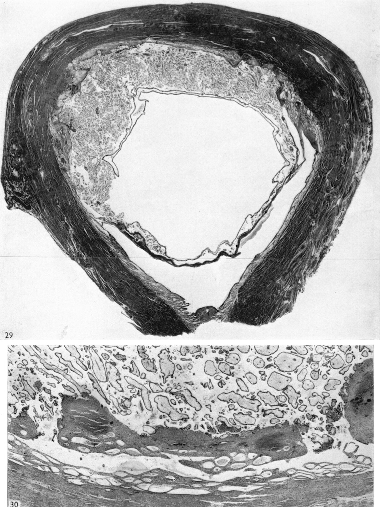
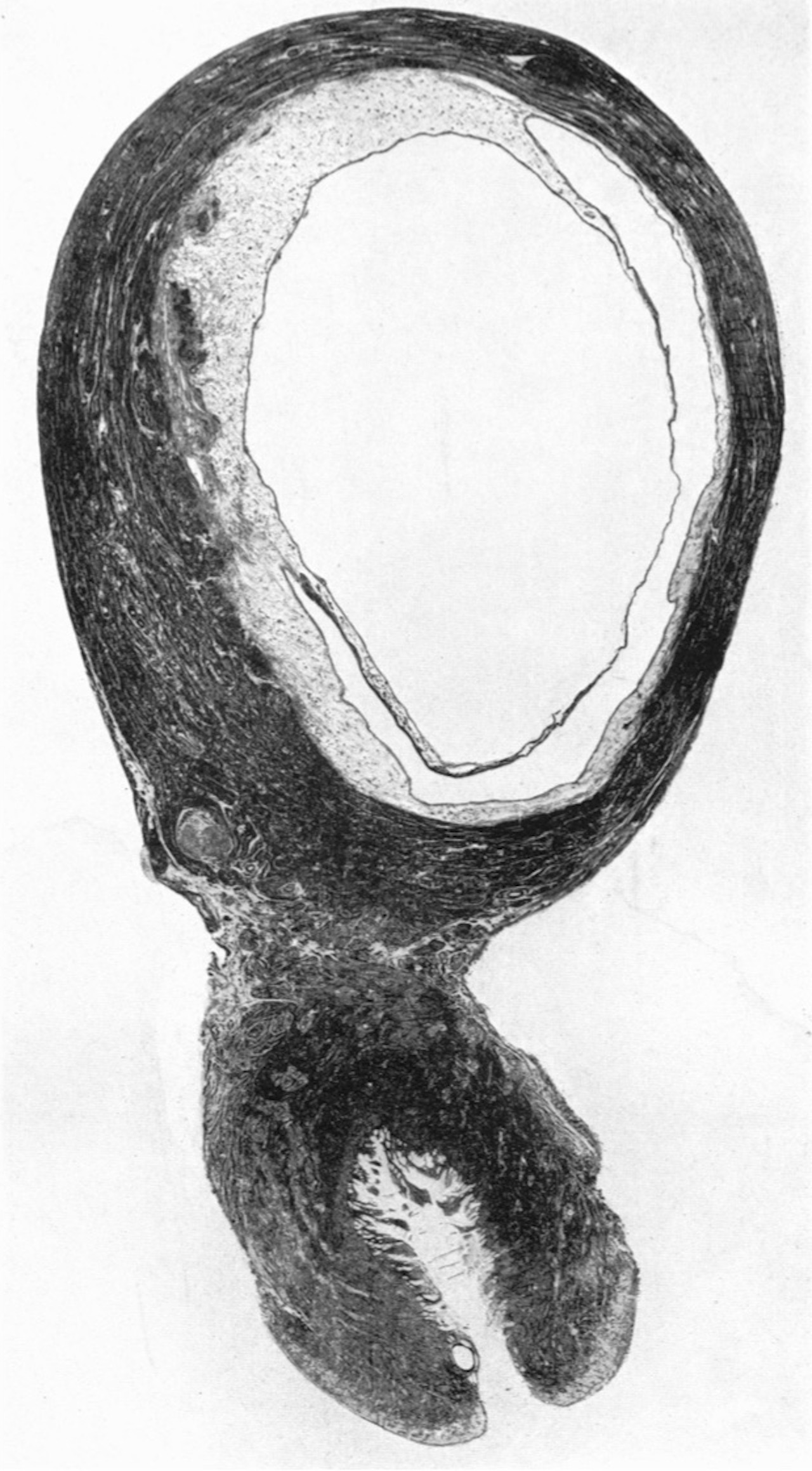
| 09:24, 14 October 2020 | Ingalls1932b fig08.jpg (file) |  |
56 KB | Z8600021 | ==Plate 100== Fig. 5. Embryo No. 210. Greatest length 15mm. Large thin-walled bleb in the midline over the rhombencephalon. Much deformity in the body generally. Fig. 6. Embryo No. 597 B. The smaller of a pair of binoval twins, greatest length 32.5mm. Very large, rather thick-walled bleb in the midline of head, just behind vertex. Extensive desquamation, malformed hands and cord. Fig. 7. Embryo No. 407. Greatest length 7 mm. Small bleb-like elevation of epithelium in the midline of the bac... | 1 |
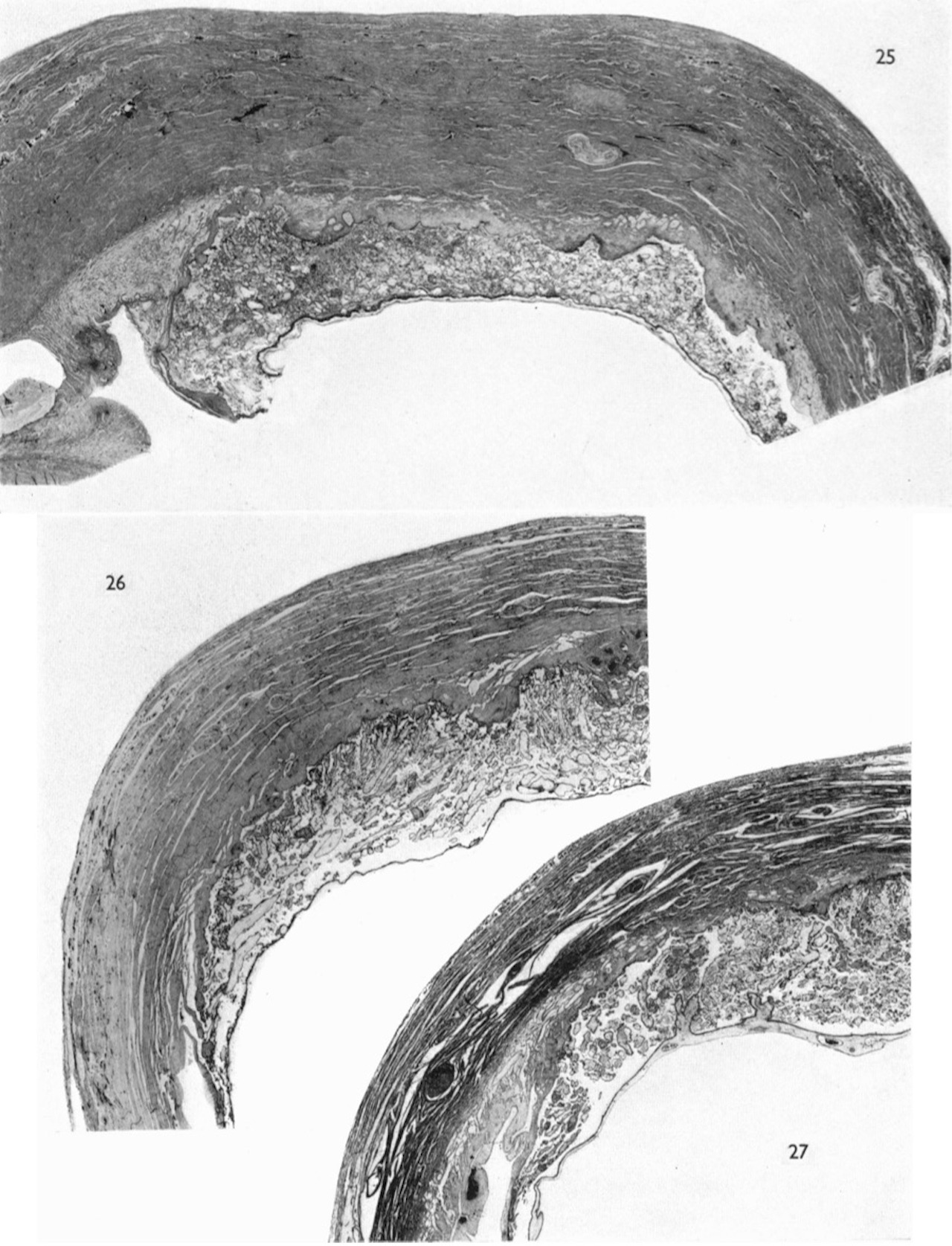
| 09:24, 14 October 2020 | Ingalls1932b fig07.jpg (file) |  |
52 KB | Z8600021 | ==Plate 100== Fig. 5. Embryo No. 210. Greatest length 15mm. Large thin-walled bleb in the midline over the rhombencephalon. Much deformity in the body generally. Fig. 6. Embryo No. 597 B. The smaller of a pair of binoval twins, greatest length 32.5mm. Very large, rather thick-walled bleb in the midline of head, just behind vertex. Extensive desquamation, malformed hands and cord. Fig. 7. Embryo No. 407. Greatest length 7 mm. Small bleb-like elevation of epithelium in the midline of the bac... | 1 |
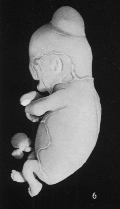
| 09:24, 14 October 2020 | Ingalls1932b fig06.jpg (file) |  |
58 KB | Z8600021 | ==Plate 100== Fig. 5. Embryo No. 210. Greatest length 15mm. Large thin-walled bleb in the midline over the rhombencephalon. Much deformity in the body generally. Fig. 6. Embryo No. 597 B. The smaller of a pair of binoval twins, greatest length 32.5mm. Very large, rather thick-walled bleb in the midline of head, just behind vertex. Extensive desquamation, malformed hands and cord. Fig. 7. Embryo No. 407. Greatest length 7 mm. Small bleb-like elevation of epithelium in the midline of the bac... | 1 |
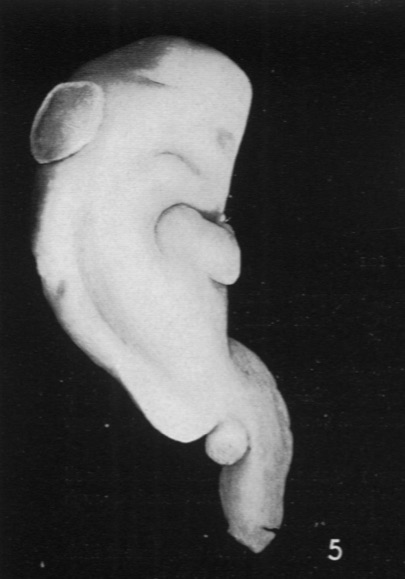
| 09:23, 14 October 2020 | Ingalls1932b fig05.jpg (file) |  |
45 KB | Z8600021 | ==Plate 100== Fig. 5. Embryo No. 210. Greatest length 15mm. Large thin-walled bleb in the midline over the rhombencephalon. Much deformity in the body generally. Fig. 6. Embryo No. 597 B. The smaller of a pair of binoval twins, greatest length 32.5mm. Very large, rather thick-walled bleb in the midline of head, just behind vertex. Extensive desquamation, malformed hands and cord. Fig. 7. Embryo No. 407. Greatest length 7 mm. Small bleb-like elevation of epithelium in the midline of the bac... | 1 |
| 09:14, 14 October 2020 | Ingalls1932b fig04.jpg (file) |  |
44 KB | Z8600021 | ==Fig. 4. Embryo No. 161== Greatest length about 12mm. Symmetrically located, transversely elongated area over the anterior part of the rhombencephalon. Embryo in very poor condition. ===Reference=== {{Ref-Ingalls1932b}} {{footer}} | 1 |
| 09:14, 14 October 2020 | Ingalls1932b fig03.jpg (file) |  |
55 KB | Z8600021 | ==Fig. 3. Embryo No. 129== Greatest length 12mm., dorsal view. Irregular, roughly circular discolored area in the anterior part of the back. Entire body badly stunted and deformed. ===Reference=== {{Ref-Ingalls1932b}} {{footer}} | 1 |
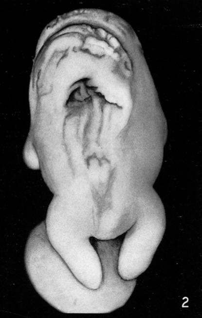
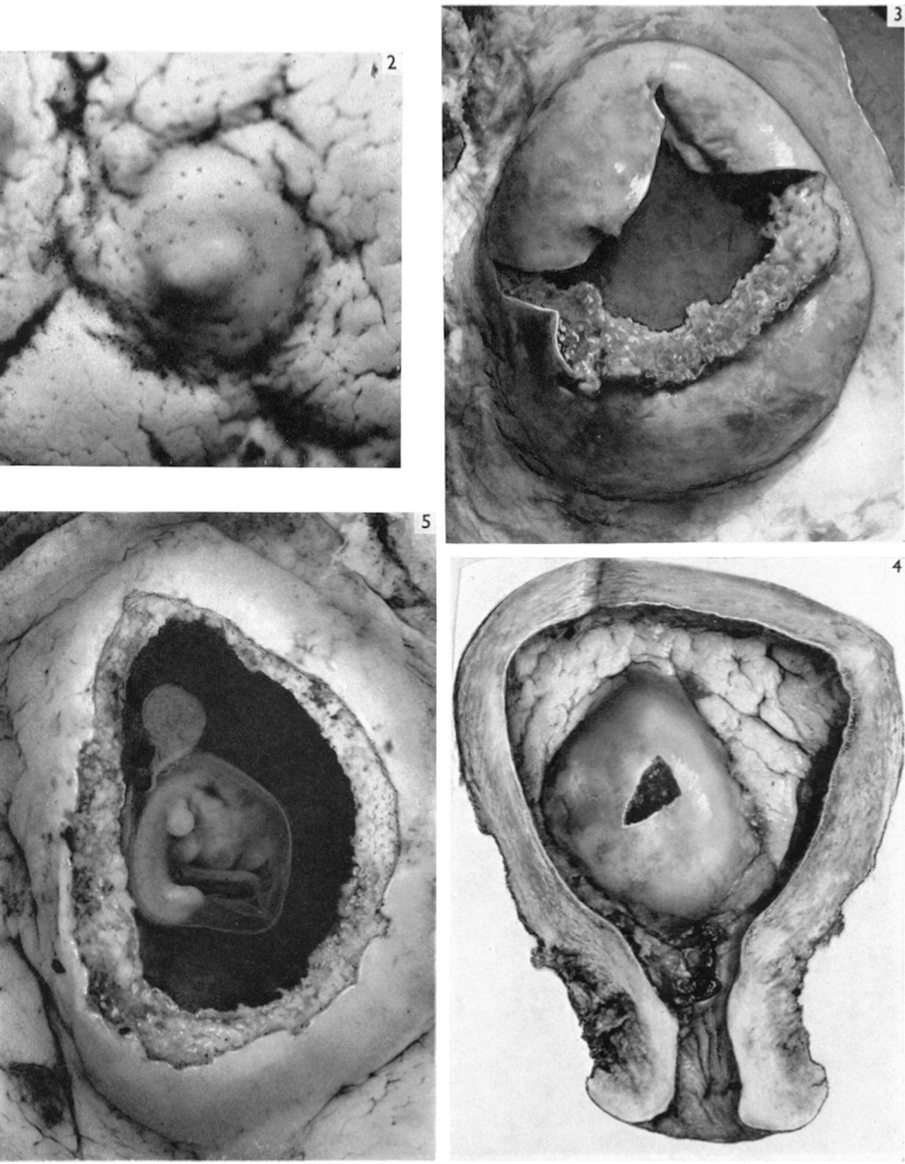
| 09:14, 14 October 2020 | Ingalls1932b fig02.jpg (file) |  |
59 KB | Z8600021 | ==Fig. 2. Embryo No. 46== Greatest length 14.5mm. The entire body is very much malformed. Almost the whole of the dorsum is markedly altered or defective. The deep triangular cavity is due to the postmortem loss of tissue. ===Reference=== {{Ref-Ingalls1932b}} {{footer}} | 1 |
| 09:13, 14 October 2020 | Ingalls1932b fig01.jpg (file) |  |
48 KB | Z8600021 | ==Fig. 1. Embryo No. 83== Greatest length about 7mm. Open neural tube in sacral region. Facial features distorted, heart exposed. ===Reference=== {{Ref-Ingalls1932b}} {{footer}} | 1 |
| 08:53, 14 October 2020 | Ingalls1932b plate101.jpg (file) |  |
174 KB | Z8600021 | ===Reference=== {{Ref-Ingalls1932b}} {{footer}} | 1 |
| 08:53, 14 October 2020 | Ingalls1932b plate100.jpg (file) |  |
197 KB | Z8600021 | ===Reference=== {{Ref-Ingalls1932b}} {{footer}} | 1 |
| 08:53, 14 October 2020 | Ingalls1932b plate99.jpg (file) |  |
192 KB | Z8600021 | ===Reference=== {{Ref-Ingalls1932b}} {{footer}} | 1 |
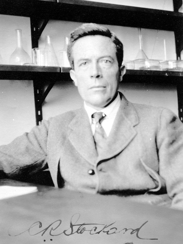
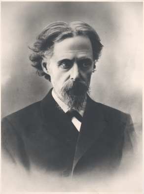
| 15:58, 28 September 2020 | Charles R. Stockard.jpg (file) |  |
393 KB | Z8600021 | Charles R. Stockard Department Of Anatomy Cornell University Medical School New York City ===Reference=== "Charles R. Stockard". History of the Marine Biological Laboratory. http://hpsrepository.asu.edu/handle/10776/3321. undated Publisher: Marine Biological Laboratory Archives ====Copyright==== Licensed as Creative Commons Attribution-NonCommercial-Share Alike 3.0 Unported. http://creativecommons.org/licenses/by-nc-sa/3.0/ https://history.archives.mbl.edu/archives/items/charles-r-stock... | 1 |
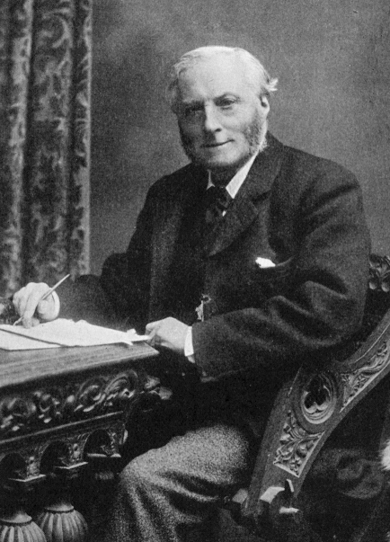
| 10:15, 20 August 2020 | Edward Long Fox.jpg (file) |  |
163 KB | Z8600021 | ===Reference=== https://www.ncbi.nlm.nih.gov/pmc/articles/PMC2511526/ | 1 |
| 11:00, 13 August 2020 | Fawcett1905 fig01.jpg (file) |  |
145 KB | Z8600021 | 2 | |
| 17:37, 12 August 2020 | Watt1915 fig01-02.jpg (file) |  |
269 KB | Z8600021 | 2 | |
| 17:26, 12 August 2020 | 1915 Transitory cavities in the corpus striatum of the human embryo.pdf (file) | 8.12 MB | Z8600021 | Category:PDF | 1 | |
| 12:04, 11 August 2020 | 1914 Nuclear masses in the lower portion of the human brain-stem.pdf (file) | 6.41 MB | Z8600021 | Carnegieinstitut191carn.pdf | 1 | |
| 09:56, 8 August 2020 | 1940 The Pineal Organ.pdf (file) | 34.62 MB | Z8600021 | Category:PDF | 1 | |
| 18:36, 6 August 2020 | HamiltonBoyd1960 fig08.jpg (file) |  |
268 KB | Z8600021 | 1 | |
| 18:36, 6 August 2020 | HamiltonBoyd1960 fig07.jpg (file) |  |
122 KB | Z8600021 | 1 | |
| 18:35, 6 August 2020 | HamiltonBoyd1960 fig06.jpg (file) |  |
97 KB | Z8600021 | 1 | |
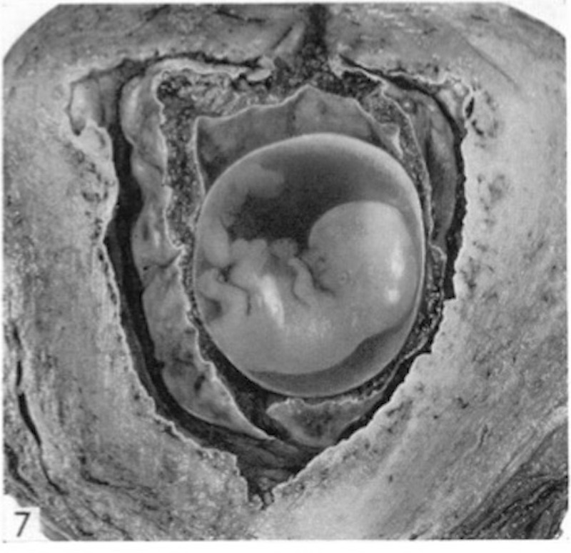
| 13:19, 6 August 2020 | HamiltonBoyd1960 fig05.jpg (file) |  |
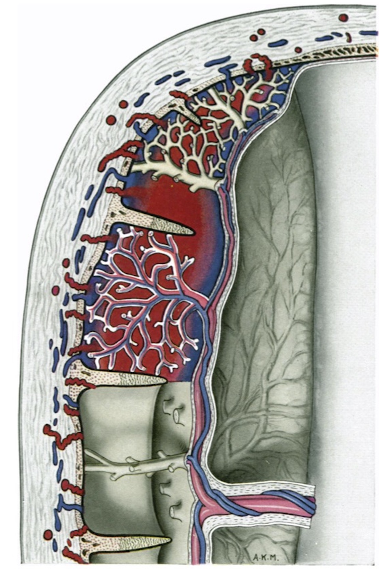
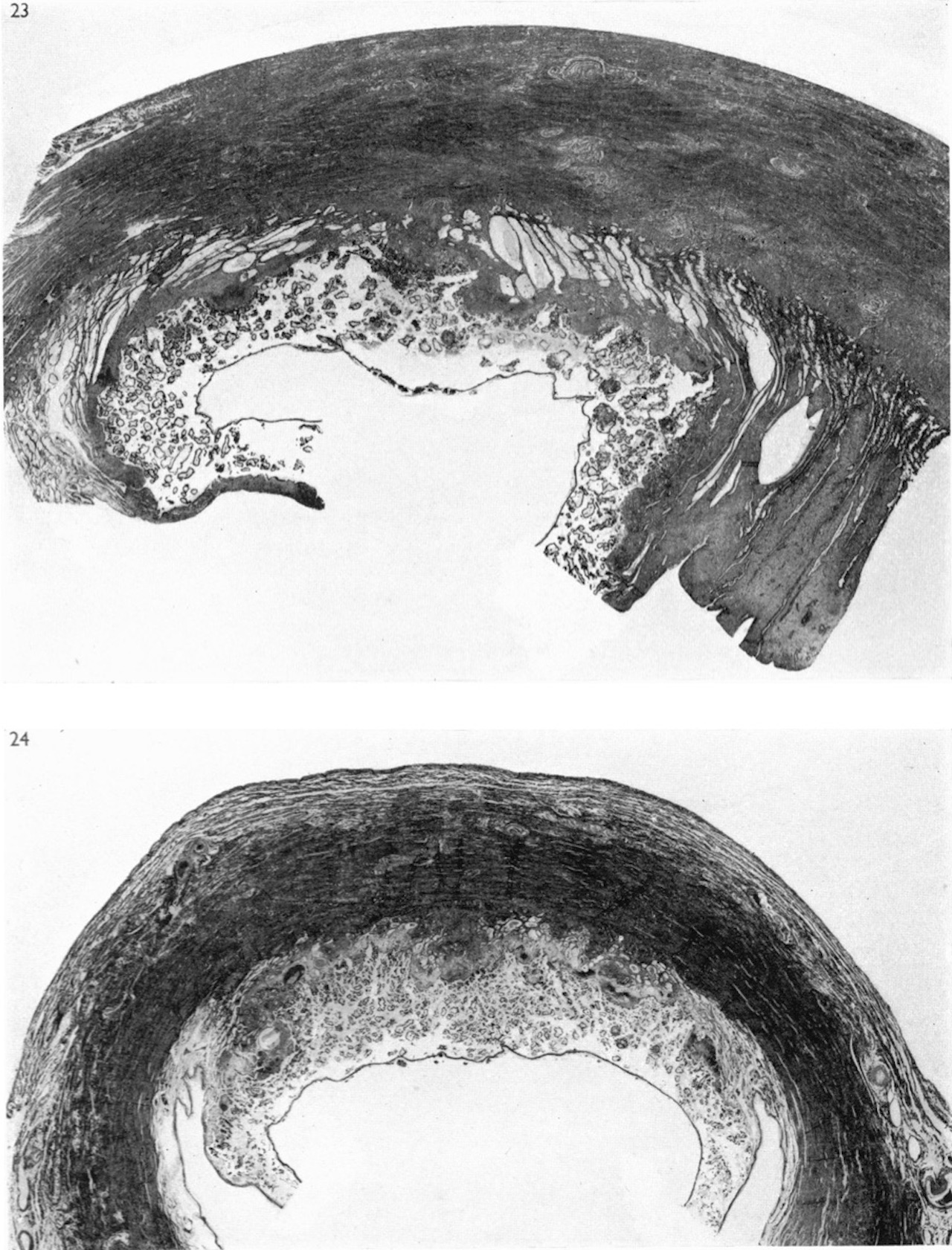
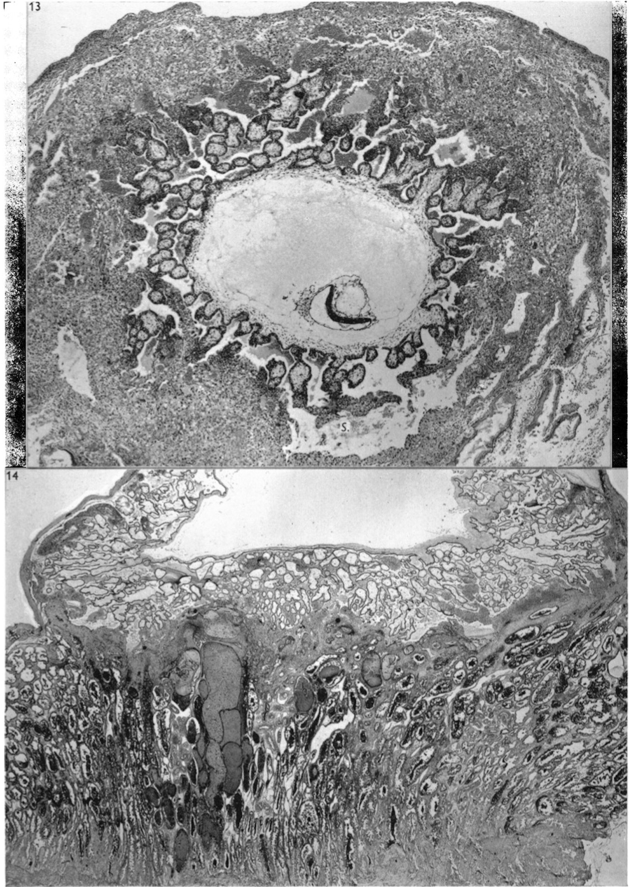
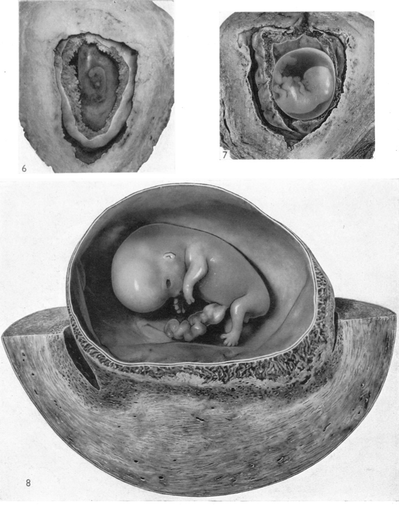
150 KB | Z8600021 | ==Fig. 5.== Photograph ( x 3-5) of the implantation site of a 10 mm. embryo (CX. 100) with the decidua capsularis removed to an extent sufficient to show the embryo, within the collapsed amnion, and the yolk sac. Note the extent of development of the chorionic villi related to the decidua capsularis. This photograph has been rotated in relation to fig. 4 to show the embryo in a suitable orientation. A section through the placenta of this specimen is illustrated in Pl. 8, fig. 23. {{Hamilto... | 1 |
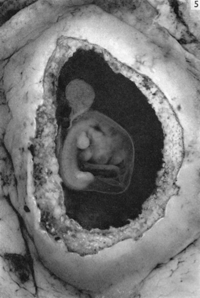
| 13:16, 6 August 2020 | HamiltonBoyd1960 fig04.jpg (file) |  |
131 KB | Z8600021 | 1 | |
| 13:13, 6 August 2020 | HamiltonBoyd1960 fig03.jpg (file) |  |
114 KB | Z8600021 | 1 | |
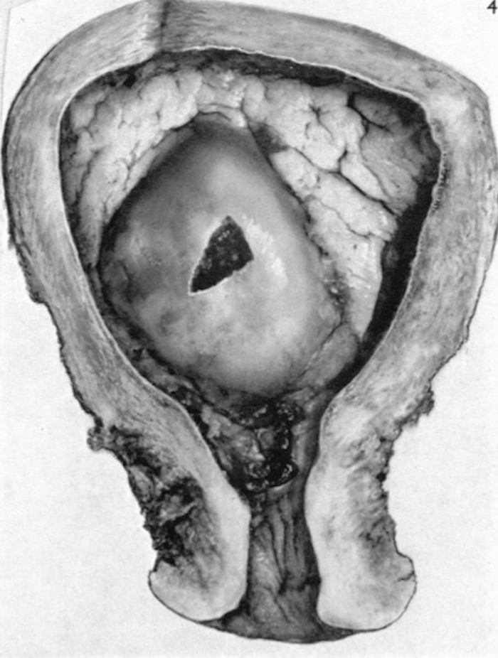
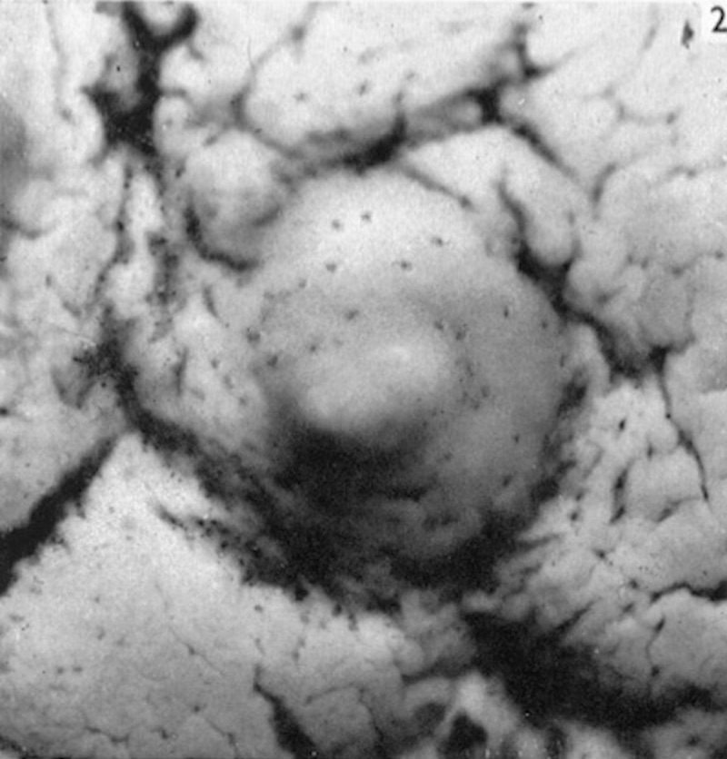
| 13:11, 6 August 2020 | HamiltonBoyd1960 fig02.jpg (file) |  |
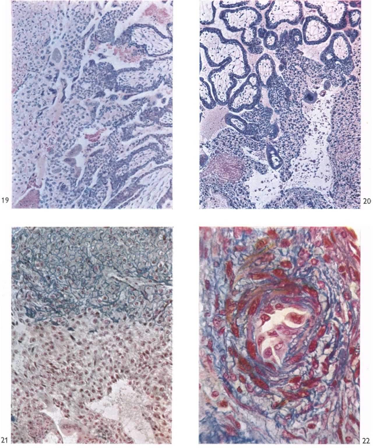
121 KB | Z8600021 | ==Fig. 2. Barnes Embryo== Photograph ( x 12) of the surface view of the implantation site of the Barnes embryo. The implantation site itself appears as a slightly elevated area of the endometrium on which openings of the uterine glands can be seen. The surface of the endometrium shows characteristic shallow and irregular furrows. | 1 |
| 12:56, 6 August 2020 | HamiltonBoyd1960 plate13.jpg (file) |  |
462 KB | Z8600021 | 2 | |
| 12:54, 6 August 2020 | HamiltonBoyd1960 plate12.jpg (file) |  |
605 KB | Z8600021 | 2 | |
| 12:51, 6 August 2020 | HamiltonBoyd1960 plate11.jpg (file) |  |
659 KB | Z8600021 | 2 | |
| 12:50, 6 August 2020 | HamiltonBoyd1960 plate10.jpg (file) |  |
499 KB | Z8600021 | 2 | |
| 12:48, 6 August 2020 | HamiltonBoyd1960 plate09.jpg (file) |  |
493 KB | Z8600021 | 2 | |
| 12:45, 6 August 2020 | HamiltonBoyd1960 plate08.jpg (file) |  |
604 KB | Z8600021 | 2 | |
| 12:41, 6 August 2020 | HamiltonBoyd1960 plate07.jpg (file) |  |
608 KB | Z8600021 | crop, adjust size | 2 |
| 12:40, 6 August 2020 | HamiltonBoyd1960 plate06.jpg (file) |  |
444 KB | Z8600021 | crop, adjust size | 2 |
| 12:39, 6 August 2020 | HamiltonBoyd1960 plate05.jpg (file) |  |
790 KB | Z8600021 | crop, adjust size | 2 |
| 12:34, 6 August 2020 | HamiltonBoyd1960 plate04.jpg (file) |  |
515 KB | Z8600021 | crop, adjust size | 2 |
| 12:30, 6 August 2020 | HamiltonBoyd1960 plate03.jpg (file) |  |
389 KB | Z8600021 | crop, adjust size | 2 |
| 12:27, 6 August 2020 | HamiltonBoyd1960 plate02.jpg (file) |  |
445 KB | Z8600021 | crop, adjust size | 2 |
| 12:21, 6 August 2020 | HamiltonBoyd1960 plate01.jpg (file) |  |
350 KB | Z8600021 | crop, adjust size | 2 |
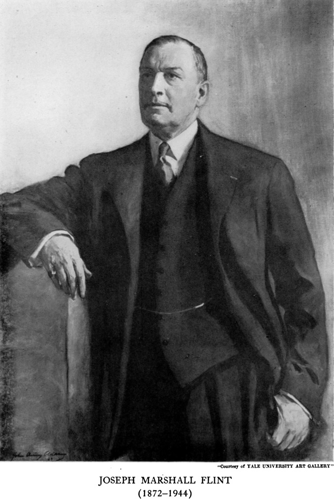
| 09:13, 6 August 2020 | Joseph Marshall Flint.jpg (file) |  |
165 KB | Z8600021 | ==Joseph Marshall Flint== | 1 |
| 11:52, 23 July 2020 | Guthrie test icon.jpg (file) | 13 KB | Z8600021 | 2 | ||
| 09:27, 22 July 2020 | Marshall1881 plate01.jpg (file) |  |
455 KB | Z8600021 | 2 | |
| 10:52, 17 July 2020 | August Rauber.jpg (file) |  |
8 KB | Z8600021 | August A. Rauber (1841-1917) was a German embryologist and anatomist. Rauber's layer is a thinned-out trophoblast membrane lying over the embryonic disk in developing carnivores and ungulates. In cattle, prevention of the loss of this polar trophoblast layer leads to ectopic domains of BRACHYURY, a gastrulation marker. :Links: {{trophoblast}} {{footer}} Category:PeopleCategory:Germany | 1 |