Human Embryology and Morphology 7
Keith, A. Human Embryology And Morphology (1921) Longmans, Green & Co.:New York.
Human Embryology and Morphology: 1 Early Ovum and Embryo | 2 Connection between Foetus and Uterus | 3 Primitive Streak Notochord and Somites | 4 Age Changes | 5 Spinal Column and Back | 6 Body Segmentation | 7 Spinal Cord | 8 Mid- and Hind-Brains | 9 Fore-Brain | 10 Fore-Brain Cerebral Vesicles | 11 Cranium | 12 Face | 13 Teeth and Mastication | 14 Nasal and Olfactory | 15 Sense OF Sight | 16 Hearing | 17 Pharynx and Neck | 18 Tongue, Thyroid and Pharynx | 19 Organs of Digestion | 20 Circulatory System | 21 Circulatory System (continued) | 22 Respiratory System | 23 Urogenital System | 24 Urogenital System (Continued) | 25 Body Wall and Pelvic Floor | 26 Limb Buds | 27 Limbs | 28 Skin and Appendages | Figures
| Historic Disclaimer - information about historic embryology pages |
|---|
| Pages where the terms "Historic" (textbooks, papers, people, recommendations) appear on this site, and sections within pages where this disclaimer appears, indicate that the content and scientific understanding are specific to the time of publication. This means that while some scientific descriptions are still accurate, the terminology and interpretation of the developmental mechanisms reflect the understanding at the time of original publication and those of the preceding periods, these terms, interpretations and recommendations may not reflect our current scientific understanding. (More? Embryology History | Historic Embryology Papers) |
Chapter VII. Central Nervous System — Differentiation of the Spinal Cord
Evolution of the Central Nervous System
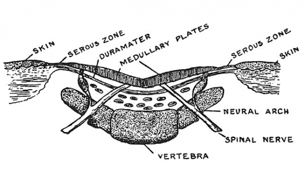
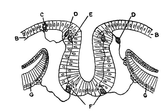
To students who are familiar with the extraordinary complexity of the central nervous system of man it must seem incredible that it arose by the specialization of an area of the ectoderm or covering of the body. It is only on such a hypothesis that we can explain the fact that the medullary plates, out of which the whole central nervous system of the body is developed, are exposed on the surface of the embryo during the greater part of the 3rd week of development. It occasionally happens that children are born, in which the medullary plates are exposed along the head and back as they are during very early embryonic life. The condition is shown in Fig. 69, and it is impossible to explain its occurrence except by supposing the medullary plates to be modified parts of the ectoderm. When, however, one remembers the condition in the lower invertebrates, such as is seen in the organization of the Hydra, the explanation becomes more acceptable. The ectodermic cells of Hydra are not only protective and secretory in function, but they also serve the purposes of nerve cells and muscle cells. One can understand how a specialization of function in the ectodermal cells may have occurred — some becoming purely contractile, others purely sensory, or secretory, or protective. In the cells of the medullary plate we see a further specialization (see Fig. 70) ; cells are specialized to connect the sensory with the contractile or muscle cells. Those connected with the sensory cells — the posterior root ganglia — arise near the lateral margins of the medullary plates ; those connected with the muscle cells arise near their mesial margins. If this hypothesis is true, then the central canal is merely an enclosed tube of ectoderm and filled with fluid, because the form of animal in which the medullary plates were evolved was a waterliving form. Dr. W. H. Gaskell has advanced the view that the central canal represents a former alimentary tube round which nerve cells have gathered. While Dr. Gaskell's hypothesis explains many facts, it leaves many more unexplained — especially the manner in which the central nervous system is developed.
Fig. 69. Diagrammatic Section across the Back of an Anencephalic Child in which the medullary plates were exposed on both head and spine.
Fig. 70. Diagram to show how the ectodermal cells of the Medullary Plates are differentiated into nerve cells or neuroblasts and supporting cells or spongioblasts. (After Prenant.) The central canal is being enclosed by upgrowth of the medullary plates. B, B, ectoderm ; C, sensory cell in ectoderm ; D, D, cells which become enclosed in posterior root ganglion ; E, E, nerve cells which connect the sensory and motor cells ; F, F, motor cells in anterior horn ; G, G, muscle plates.
Formation of the Central Canal
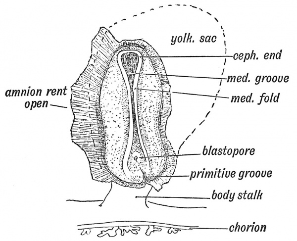
The medullary plates of ectoderm, which form the spinal cord and brain, rise up, meet, and enclose a canal — the central canal of the spinal cord and brain (Fig. 71). The lips of the medullary plates meet alnd fuse together in the cervical region first, the process of union spreading forwards and backwards, the last parts to be enclosed being the cephalic and caudal extremities. The opening at the anterior extremity — the neuropore — and the posterior or caudopore close towards the end of the 4th week, the neuropore closing first. The optic vesicles begin to grow out from the medullary plates before these have united to enclose the cavity of the fore-brain. It will be thus seen that the optic vesicle, which becomes the retina and optic nerve, is developed as a part of the medullary plate.
Fig. 71. Medullary Folds uniting to form the Neural Tube in a Human Embryo in the 3rd week of development. (After Graf Spee.)
|
|
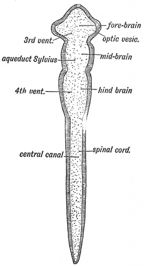
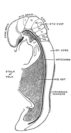
| Fig. 72. Diagram of the Four Primary Divisions of the Neural Tube. | Fig. 73. Lateral View of the Central Nerve System of a Human Embryo of the 4th week — 2.6 mm. long. (Dr. Low.) |
Division of the Neural Canal (Figs. 72, 73).— At the end of the 4th week the neural tube is divided into four parts. They are :
- An anterior dilatation, the fore-brain, which forms the 3rd and lateral ventricles and their walls.
- The mid-brain, which becomes transformed into the aqueduct of Sylvius, corpora quadrigemina and crura cerebri.
- the hind-brain, the basis of the 4th ventricle, pons, cerebellum and medulla.
- The central canal and spinal cord.
The Spinal Cord
The Spinal Cord at first extends throughout the whole length of the spinal column. At the end of the 3rd month the spinal column and canal grow more rapidly than the cord, and at birth its lower end has become withdrawn to the level of the 3rd lumbar vertebra.[1] By the third year it usually terminates opposite the disc between the 1st and 2nd lumbar vertebrae, but it may stop at the lower border of the 2nd lumbar or rise as high as the middle of the 12th dorsal vertebra.[2] The results of this inequality of growth are : (1) The roots of the lumbar and sacral nerves become enormously elongated, forming the cauda equina ; all the nerves are more or less drawn up, except the 1st and 2nd cervical ; the origins of the lower cervical nerves are drawn up 2 vertebrae (as indicated by the position of their spines) ; the upper dorsal, 3 ; the lower dorsal, 4 ; the lower lumbar, 5 ; the coccygeal, 10. These statistics represent a broad expression of the observations made by Professor R. W. Reid.
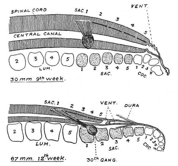
(2) The caudal part of the spinal cord is the last part of the neural tube to be formed (see Fig. 63). Its fate has been recently investigated by Professor Streeter.[3] Even in the 9th week (Fig. 73, A) the caudal segment is still represented over the coccyx, ending in a subcutaneous vesicle, but already retrogression has set in, the coccygeal ganglia have disappeared and the neural canal, immediately distal to the origin of the 5th pair of sacral nerves, is becoming dilated to form the terminal ventricle. By the 12th week (Fig. 74), when retraction has set in, we see that the caudal segment has become differentiated into a distal or extradural part, which is drawn out to form the coccygeal thread, while the intradural part is being stretched and will become the filum terminale.
Fig. 73, A. Showing the differentiation of the terminal part of the neural tube into the coccygeal thread and fllum terminale. (Streeter.)
Differentiation of the Spinal Cord
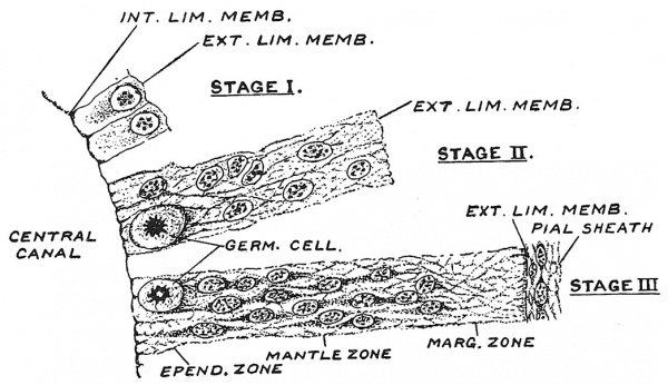
As the neural plate is folded in towards the end of the 3rd week, the single layer of columnar epithelium of which it is composed is already undergoing certain changes. Three stages in its differentiation are shown in Fig. 74 ; in Stage I., the single layer of ill-defined columnar cells is shown ; the bases of the cells are directed towards the central canal, resting on a delicate internal limiting membrane ; their outer ends, appearing on the surface of the neural tube, are bounded by the external limiting membrane. In Stage II. there has been an active proliferation of the cells and an increase in the thickness of the neural wall ; the cell bodies have fused to form a cytoplasmic syncytium, in which the nuclei are spread between the inner and outer limiting membrane. In Stage III. (Fig. 74), which is reached about the close of the 4th week, the wall has made a further increase in thickness ; in the common C3rtoplasm a fibrillar meshwork — a myelospongium — has been laid down ; three zones can be distinguislied, a middle or mantle zone in which most of the nuclei are contained, which will become the grey substance of the cord ; an outer or marginal zone made up of myelospongium into which the fibre-tracts of the cord will grow ; an inner or ependymal zone, not distinctly demarcated, but characterized by the presence of actively dividing large nuclei — germinal cells. The nuclei, with their surrounding protoplasm, are becoming differentiated into neuroblasts — the producers of nerve cells, and neuroglial or supporting cells. The neuroglial fibres are laid down in the cell-protoplasm of neuroglial cells. The ependymal cells which line the central canal, are derived from the inner zone.
Fig. 74. Three stages in the early differentiation of the wall of the spinal neural tube. Stage I., single layer of ill-differentiated columnar epithelium ; Stage II., in which the single layer has been transformed into a nucleated syncytium ; Stage III., in which three zones begin to be apparent. After George Streeter (1873-1948)
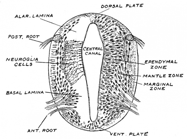
Fig. 75. Section across the developing Spinal Cord at the beginning of the 5th week. After Wilhelm His (1831-1904)
A section across the embryonic spinal cord at the beginning of the 5th week (Fig. 75) brings out certain instructive features : the central canal is coffin-shaped in section ; the epithelium in its roof and floor — forming the roof and floor plates — increases but slightly in thickness ; its side walls, walls, or lateral plates, which become the cord or medulla, are indistinctly separated into a ventral part or basal lamina, from which the anterior root fibres emerge and winch will have to do with motor functions and a dorsal part — the alar lamina, into which the fibres of the posterior root grow and which will have to do with sensory functions. The three zones in each lateral plate are distinct ; the inner or ependymal zone increases in breadth as it is followed from the floor plate to the roof plate, whereas the middle or mantle zone does the opposite ; it diminishes as it passes into the alar lamina. In the ventral part of the middle zone the anterior grey column or horn is quite apparent, whereas the posterior horn is just beginning to form. We must suppose that the inner or ependymal zone is one of production or proliferation and that its cells are becoming differentiated and added to the middle zone. The anterior or motor column is demarcated before the posterior or sensory horn. In each lateral plate the neuroblasts become grouped thus from anterior to posterior horn : (1) somatic motor, (2) splanchnic motor (both in the basal lamina), (3) splanchnic sensory, (4) somatic sensory (both in the alar lamina). This order of grouping holds true from end to end of the neural tube. The fibres from the cells in the anterior horn begin to emerge as the anterior root in the latter part of the 4th week ; the processes from the ganglion cells of the posterior root commence to enter the marginal zone of the alar lamina at the same time. As is diagrammatically represented in Fig. 75 neuroglial fibres pass from the lining ependyma of the central canal to the external limiting membrane.
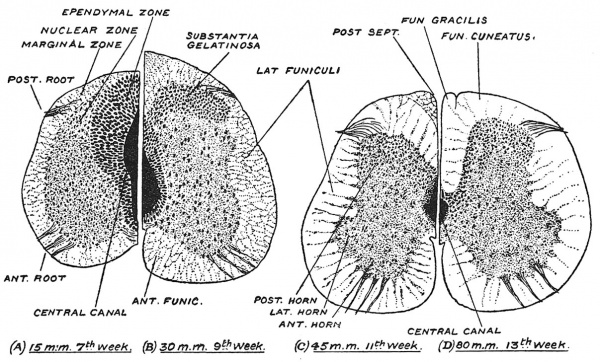
Fig. 76. Showing the progressive differentiation of the spinal cord during the second and third months of development. (After Streeter.)
The developmental changes in the cord during the 2nd and 3rd months are set out in a semi-diagrammatic manner in Fig. 76, A, B, C, D. We may centre our attention first on three structures — the central canal, the inner or ependymal zone and the posterior median septum, for all three are closely correlated. In the 7th week (Fig. 76, A) the central canal still retains its coffin-shaped section, the ependymal zone is still extensive, and although the roof plate has thickened there is still no posterior median septum, for the posterior funiculi or conducting tracts (f . gracilis and f. cuneatus) have scarcely appeared in the dorsal marginal zone. In the 9th week changes are in progress : the dorsal part of the central canal is being obliterated by the apposition of the lateral plates (Fig. 76, B) ; the ependymal zone is reduced ; the posterior funiculi are being formed in the dorsal marginal zone and the posterior-median septum is formed between the right and left posterior funiculi developing on each side of the original roof plate. In the 11th week the central canal and ependymal zone are further reduced ; the posterior-median septum has increased in depth owing to the rapid growth of the posterior funiculi ; the middle or mantle zone is now differentiated into the anterior and posterior columns of grey matter. In the 13th week the adult condition is reached ; the cord is reduced to its final size, the ependymal zone now forms merely a lining to the canal ; the anterior and posterior horns, with their various groups of nerve cells, are reaching their final form, while in the marginal zone the great connecting and association tracts of white matter have arisen or are arising. There is now a deep posterior median septum and an open anterior median fissure, formed during the development of the ventral funiculi in the anterior part of the marginal zone.
Spinal Tracts
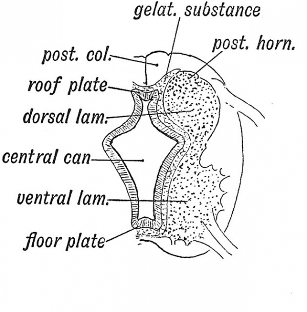
With the formation of the posterior columns, the grey matter of the dorsal laminae, at first united by the roof plate, becomes widely separated to form the posterior horns (Fig. 77). At the same time part of the gelatinous tissue of the inner zone is separated to form a cap on the posterior horns (Fig. 76). In the gelatinous tissue congenital cysts may occur. The columnar cells which line the central canal are ciliated. Thus by the end of the 3rd month the nerve cells have taken up their permanent stations in the grey columns of the spinal cord. The cells which have to do with the reception and transmission of sensory messages are situated in the posterior root ganglia ; those which have to do with the dispatch of motor impulses are situated in the anterior and lateral horns ; the remainder may be regarded as intercalated or shunt cells, and are concerned in linking up or associating the afferent and efferent systems and centres. The marginal zone provides a basis into which the nerve processes — the axons which are to connect neuron with neuron and centre with centre — may grow and reach their destinations. It is a remarkable fact that the lower we go in the vertebrate scale the more automatic or independent do the nerve centres of the spinal cord become ; the higher we go in the scale the more they become dominated by and dependent upon nerve centres situated in the hind-brain, mid-brain and fore-brain. Hence we are prepared to find that the first tracts of nerve fibres which appear in the marginal zone are those which link together the nerve centres in the spinal cord itself. At the end of the first month the fibres of the posterior root have entered the marginal zone on the dorsal side of the cord, and have thus formed the rudiment of the posterior funiculi ; these effect connections with receptive nuclei in the posterior horns. At the same time fibres which associate neighbouring or allied nuclei or centres of the cord aj)pear in the marginal zone of the lateral and anterior parts of the cord. These may be described as inter-segniental tracts.
Later, in the 3rd month, commences the growth of fibres within the antero-lateral marginal zone of (1) tracts which, arising from cells in the cord, are to end in hind-brain, mid-brain and fore-brain, and thus supply these higher centres with afferent impulses which are reaching the spinal centres ; (2) tracts which, commencing in the hind and mid-brain, grow down to permit the higher centres to influence the lower centres in the cord. Lastly, in the 5th month, the pallio-spinal or pyramidal tracts commence to develop. The pyramidal tracts (crossed and direct) grow down from the cells of the motor cortex. They are not medullated until soon after birth. The pyramidal tracts are the means by which the brain controls the motor cells of the cord. In man these tracts are remarkable, not only for their great size, but also that in addition to the crossed lateral tract, which is present in all mammals, there is also an anterior or direct tract. The anterior tract appears to be a recently evolved system ; it is extremely variable in size. The only other animals which possess it are those nearest allies of man — the great anthropoid apes.
Fig. 77. Diagrammatic Section of the developing Spinal Cord to show (1) the Roof and Floor Plates ; (2) the Dorsal (alar) and Ventral (basal) Laminae ; (3) the Gelatinous Tissue between the Middle and Inner Zones.
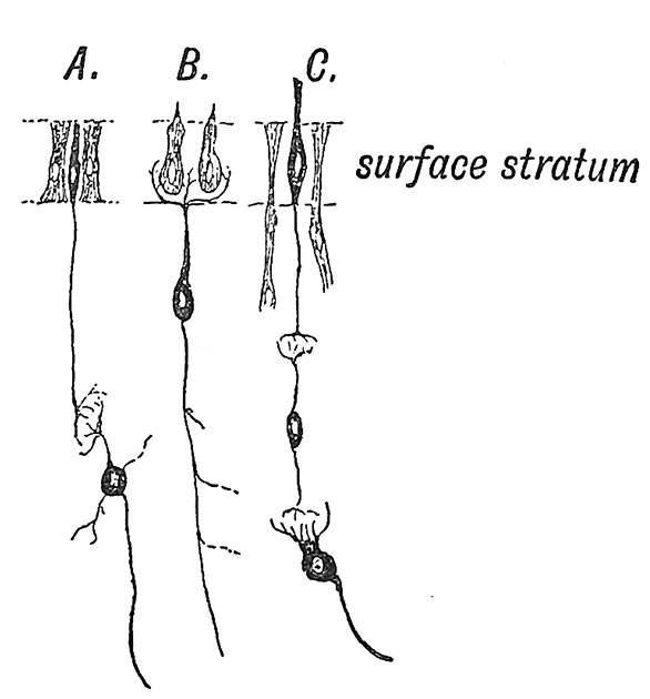
Fig. 78. Showing Transformation of Cells of the Ectoderm to Sense Epithelium, Nerve Cells and Supporting Cells, in A, the Olfactory Plate, B, the Otocyst, C, the Retinal Layer of Optic Cup.
Myelinization
The myelinization[4] — the formation of medullary sheaths for the fibres of nerve tracts — commences about the 4:th month and is not really finished until the age of puberty is reached. The oldest tracts — the ones which are first required to carry messages — are the earliest to be medullated. The process begins in the oldest part of the fibre — the part nearest the parent nerve-cell — and spreads towards the growing tip. The great nerve tracts are ensheathed at different dates ; hence it is possible to distinguish and unravel one tract from another during the period of development.
Segments of the Spinal Cord
To that part of the neural tube and neural crest which corresponds in position to a primitive body-segment, the name of Neuromere is given. From the cells of a neuromere are produced the posterior and anterior root of a spinal nerve on each side. The extent of each neuromere is thus marked out by the attachments of its nerve roots. At no time are the medullary plates divided into embryological segments in the same sense as the mesoderm is divided, although the neural tube probably did arise from the fusion of a series of neuromeres or ganglia, each presiding over a definite segment of the body, subsequent evolutionary changes have led to their fusion. Dr. Watt observed in a human embryo in which there were 18 body-somites, that 11 segments were to be noted in the spinal cord. These changes are related to the combination of the various segments and systems in carrying out the functions of the body. The cervical and lumbar enlargements of the cord appear in the 4th month. They contain the neuroblasts connected with the body-segments which gave rise to the upper and lower extremities. The neuroblasts are arranged, not according to the original neural segments, but rather in relationship to the movements of the limb. The group representing the hand movements lie behind (distal to) those representing movements of the forearm.
Origin of the Medullary Plates and Nerve Cells
The medullary plates like the olfactory plates which give rise to the sense-epithelium of the nose, the otocyst from which the auditory organ is developed, and the retina are derived from the ectodermal covering of the embryo. The olfactory plate retains to the greatest extent the features of the ectoderm (Fig. 78, A). Its cells are of three kinds : (1) protective, (2) secretory, (3) sensory, the latter being essentially surface nerve cells in nature. A process or axis cylinder is produced from each sense cell ; from its opposite extremity a sensory process is produced (Fig. 78, A). In worms, sense epithelial cells sink beneath the protective and secretory cells, the sensory process being drawn out to form a fibre. In the otocyst, the sensory cells produce no axis-cylinder process, but a ganglionic cell — produced from the ectoderm through the neural crest — comes into connection with it (Fig. 78, B). From the ganglionic cells are produced (1) a chief process or axis cylinder ; (2) a branching process or processes — dendrites — from the opposite pole, which end in an arborescence round the sensory cells. To a nerve cell and all the processes developed from it the name of Neuron is given. In the retina, as in the olfactory plate, three types of cells are seen : (1) protective or supporting which form the fibres of Miiller, (2) secretory over the ciliary processes, (3) the sensory cells, which produce an axis cylinder on one side, and a rod or cone on the other (Fig. 78, C). Further, by a process of division, bipolar and ganglionic cells are produced from the retinal sense cells. In the medullary plates of the spinal cord the representatives of the original ectoderm form the ependymal and neuroglial cells, the first of which may be regarded as both secretory and supporting ; the neuroblasts arise by a process of division from the primary ectodermal cells. Each neuroblast gives rise to a neuron. Their axons or axis cylinders are in many cases two feet or more in length ; for instance, the motor and sensory fibres which pass from the lumbar enlargement to the muscles and skin of the foot. The nerve cells in the basal laminae are peculiar in that their axis cylinders end on muscle cells.
Malformations of the Neural Canal
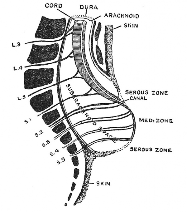
The fact that children are occasionally born with the medullary plates open and exposed on the head and back has already been mentioned (see Fig. 69).[5] Total Rachischisis, as the condition is named, is rare ; it is much more usual to find only one part of the neural tube open — either the anterior or cephalic part, giving the condition known as Anencepiialy — absence of brain, or the posterior or lumbo-sacral part, giving the condition known as cystic spina bifida. The latter condition is shown in Fig. 79. As the spinal cord is followed down, it is seen to enter a cystic structure formed by a dilatation of the subarachnoid space, across which the roots of the lumbo-sacral nerves pass. The projecting dome of the cyst is formed by the expanded medullary plates ; hence the spinal cord appears to end on the wall of the cyst, and spinal nerves to actually arise from it. The lumbo-sacral parts of the neural tube and of the spine have never been enclosed ; the cerebro-spinal fluid collects in the subarachnoid space, and the unresisting medullary plates are raised up to form part of the wall of a cystic tumour. Another form of pathological dilatation may appear after the neural tube is completely closed. In chicks hatched at abnormal temperatures fluid may collect in certain parts of the tube, thus dilating it and giving rise to cystic conditions.
Fig. 79. Vertical Section of the Lumbar Region to show the arrangement of parts in a typical case of cystic spina bifida.
Membranes and Vessels of the Cord
When the neural tube is enclosed towards the end of the 3rd week by the upgrowth of mesoderm in the medullary folds, mesenciiymal cells become applied to the neural tube. They form the primary sheath of the neural tube (Fig. 74, C). The sheath receives a vascular supply from each dorsal branch of the segmental arteries and veins. Branches of the vessels perforate the nerve tissue, and thus a vascular mesodermal element is added to the ectodermal neural laminae. By the middle of the second month the primary sheath has become cleft into an inner or pial layer, and an outer or arachno-dural layer. The cleft becomes the subarachnoid space, which is apparently of the nature of a lymphatic space (see p. 90).
Development of Nerves[6]
In lower fishes Kupffer found that nerve fibres were formed by the union of a chain of cells — probably ectodermal in origin, and many suppose that the nerve fibres of all vertebrates are formed in this manner, the nuclei of the chain-cells becoming the nuclei of the neurolemma. On the other hand His and Kolliker concluded that every nerve fibre is produced as a continuous outgrowth from one nerve cell, and that the cells of the sheath are mesodermal in origin. It is possible that both interpretations of the apjDearance presented by developing nerves are right, and that one kind of nerve fibre is produced in the first manner and another kind in the second manner. The opinion generally held at the present time is that an axis cylinder is the product of one nerve cell or neuroblast, and that the cells which surround the growing fibres and form their sheaths are derived from the neural crests, and are therefore ectodermal in origin. It is maintained by Dr. John Cameron that these surrounding cells assist in the deposition of an achromatic substance at the growing points of nerve fibres {Journ. Anat. and Physiol. 1906, vol. 41, p. 8). Dr. Ross Harrison found that when small parts of the medullary plates of tadpoles were transplanted or maintained alive in artificial media the outgrowth of the neurons as processes from single cells could be witnessed {Anat. Record, 1908, vol. 2, Nos. 9, 10).
Another theory receives support from Graham Kerr's recent investigations on Lepidosiren, viz. that nerve fibres are formed by the stretching of protoplasmic connections which originally exist between nerve and muscle cells.
- ↑ G. L. Streeter, Amer. Journ. Anat. 1919, vol. 25, p. 1.
- ↑ R. E. McCotter, Anat. Rec. 1916, vol. 10, p. 559.
- ↑ G. L. Streeter, Amer. Journ. Anat. 1919, vol. 25, p. 1.
- ↑ Florence R. Sabin, Amer. Journ. Anat. 1911, vol. 11, p. 113 (Model of Tracts medullated at Birth).
- ↑ J. P. Good, Journ. Anat. 1912, vol. 46, p. 391 ; J. Voigt, Anat. Hefte, 1906, vol. 30, p. 393 ; W. M. Baldwin, Anat. Record, 1915, vol. 9, p. 365 ; Theodora Wheeler, Contrib. to Embryology, 1920, vol. 9, p. 95 ; E. J. Carey, Anat. Record, 1919, vol. 16, p. 45.
- ↑ Papers on histogenesis of nerves : R. G. Harrison, Amer. Journ. Anat. 1906, vol. 5, p. 121 ; W. H. Lewis, Amer. Journ. Anat. 1906, vol. 6, p. 461 ; J. Cameron, Journ. Anat. and Physiol. 1907, vol. 41, p. 8 ; Prof. T. H. Bryce, Quain's Anatomy, 1908, vol. 1, p. 94.
| Historic Disclaimer - information about historic embryology pages |
|---|
| Pages where the terms "Historic" (textbooks, papers, people, recommendations) appear on this site, and sections within pages where this disclaimer appears, indicate that the content and scientific understanding are specific to the time of publication. This means that while some scientific descriptions are still accurate, the terminology and interpretation of the developmental mechanisms reflect the understanding at the time of original publication and those of the preceding periods, these terms, interpretations and recommendations may not reflect our current scientific understanding. (More? Embryology History | Historic Embryology Papers) |
Human Embryology and Morphology: 1 Early Ovum and Embryo | 2 Connection between Foetus and Uterus | 3 Primitive Streak Notochord and Somites | 4 Age Changes | 5 Spinal Column and Back | 6 Body Segmentation | 7 Spinal Cord | 8 Mid- and Hind-Brains | 9 Fore-Brain | 10 Fore-Brain Cerebral Vesicles | 11 Cranium | 12 Face | 13 Teeth and Mastication | 14 Nasal and Olfactory | 15 Sense OF Sight | 16 Hearing | 17 Pharynx and Neck | 18 Tongue, Thyroid and Pharynx | 19 Organs of Digestion | 20 Circulatory System | 21 Circulatory System (continued) | 22 Respiratory System | 23 Urogenital System | 24 Urogenital System (Continued) | 25 Body Wall and Pelvic Floor | 26 Limb Buds | 27 Limbs | 28 Skin and Appendages | Figures

