Human Embryology and Morphology 5
Keith, A. Human Embryology And Morphology (1921) Longmans, Green & Co.:New York.
Human Embryology and Morphology: 1 Early Ovum and Embryo | 2 Connection between Foetus and Uterus | 3 Primitive Streak Notochord and Somites | 4 Age Changes | 5 Spinal Column and Back | 6 Body Segmentation | 7 Spinal Cord | 8 Mid- and Hind-Brains | 9 Fore-Brain | 10 Fore-Brain Cerebral Vesicles | 11 Cranium | 12 Face | 13 Teeth and Mastication | 14 Nasal and Olfactory | 15 Sense OF Sight | 16 Hearing | 17 Pharynx and Neck | 18 Tongue, Thyroid and Pharynx | 19 Organs of Digestion | 20 Circulatory System | 21 Circulatory System (continued) | 22 Respiratory System | 23 Urogenital System | 24 Urogenital System (Continued) | 25 Body Wall and Pelvic Floor | 26 Limb Buds | 27 Limbs | 28 Skin and Appendages | Figures
| Historic Disclaimer - information about historic embryology pages |
|---|
| Pages where the terms "Historic" (textbooks, papers, people, recommendations) appear on this site, and sections within pages where this disclaimer appears, indicate that the content and scientific understanding are specific to the time of publication. This means that while some scientific descriptions are still accurate, the terminology and interpretation of the developmental mechanisms reflect the understanding at the time of original publication and those of the preceding periods, these terms, interpretations and recommendations may not reflect our current scientific understanding. (More? Embryology History | Historic Embryology Papers) |
Chapter V. The Spinal Column and Back
Stages in the Development of the Spinal Column
In previous chapters the main facts relating to the development of the human body during the first and second months have been briefly sketched. We now turn to the consideration of particular parts of the human body, and naturally take up first the vertebral column — the main axis of the body. The most primitive form of axial support — the notochord — appears in the embryo during the third week. In amphioxus the notochord forms a permanent structure ; in all vertebrate animals it is replaced by a segmented or vertebral axis. In the evolution of the spinal column three stages are recognized : (1) one in which the skeletal segments were composed of cellular or mesenchymatous tissue ; (2) a cartilaginous stage, in which the cells of the mesenchyme (see p. 40) become modified into cartilage-forming or chondrogenous cells ; (3) a final stage where the cartilage is replaced by bone. In the human embryo we see those three stages appear in succession ; at the beginning of the second month the membranous foundation of the vertebra is being laid down ; in the middle of that month the cartilaginous change has commenced ; by the beginning of the third month ossification has commenced. In only certain groups of fishes is the cartilaginous stage a permanent one.
Stages in the Evolution of the Human Spinal Column
We have already seen that the vertebral column and its muscles appear first as a great flexible scull for driving the animal forwards (p. 41), but in nearly all mammals the vertebral column comes to serve as a horizontal axis or arch, which is supported on the fore and hind limbs. In a small group, however, which includes the anthropoid apes and man, the spinal column no longer forms a horizontal but a vertical axis or column. These higher primates are upright or orthograde when they move, in contradistinction to the ordinary four-footed mammals which are pronograde. There is no doubt that the orthograde posture was evolved from the pronograde. Although the anthropoid apes are orthograde, yet they use their arms in locomotion, to assist their lower extremities in supporting the weight of their bodies. Man is also orthograde, but he differs from the anthropoids in supporting the weight of his body entirely on his lower extremities. Hence we find that the spinal column of man, although similar to that of the anthropoids, shows many peculiar adaptations to his manner of locomotion. These adaptations become especially manifest as the child learns to walk, and are best realized by a survey of the pyramids and curves of the spine.
The Pyramids of the Spine
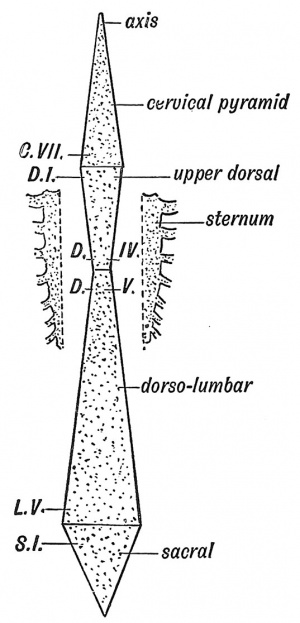
The spine, when viewed from the front, is seen to be made up of four pyramids : (1) Cervical ; (2) upper dorsal ; (3) dorso-lumbar ; (4) sacrococcygeal (Fig. 47). The bases of the two upper pyramids meet at the disc between the 7th cervical and 1st dorsal vertebrae ; the bases of the lower two at the disc between the 5th lumbar and 1st sacral vertebrae. The apices of the two middle pyramids meet at the disc between the 4th and 5th dorsal vertebrae, which have therefore the narrowest bodies of the vertebral series. The narrowing in the upper dorsal region is due to the fact that the weight of the upper half of the trunk is partly borne by, and transmitted to, the lower dorsal region by the sternum and ribs which thus relieve the spine to some extent (Fig. 47). At the sacrum the weight is transferred to the pelvis and lower limbs, hence the rapid diminution of the sacrum and coccyx. A well-marked thickening or bar in each ilium runs from the auricular surface to the acetabulum and transmits the weight to the femora.
The Curves of the Spinal Column
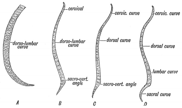
There is only one curve — an anterior concavity — until the 3rd month (Fig. 48, A). About the beginning of the 4th month the sacro-vertebral angle forms between the lumbar and sacral regions (Fig. 48, B). At birth the cervical and sacral curves have appeared, but the sacral not to a pronounced extent (Fig. 48, C). The lumbar curve appears as the child learns to walk. It is produced to allow the body to be brought vertically over the lower extremities. The sacral and cervical curves also become at that time more marked (Fig. 48, D). The dorsal curvature and the sacro-vertebral angle are the primitive curves and are present in all mammals. The others are adaptations to the upright posture. The lumbar curve is most pronounced in the highly civilized races.
Fig. 48. Diagram of the Curves of the Spinal Column. A. At the 6th week of foetal life. B. At the 4th month of foetal life C. Curves present present at Birth. D. Curves in the Adult.
Proportion of Cartilage and Bone
The intervertebral discs form one-third of the total height of the spine ; the proportion of cartilage is greater in the lumbar than in the dorsal region and greater in the dorsal than in the cervical. The lumbar and cervical curvatures are due chiefly to the shape of the discs (H. Morris). In the lumbar region, which is convex forwards, only the lower three vertebrae are deeper in front than behind. This is true only for the higher races of mankind, for as Cunningham has shown, in lower races, as in the gorilla, only the last lumbar vertebra is deeper in front than behind, and thus helps to maintain the lumbar curvature.
Unstable Regions of the Spine
In about 90 % of men there are 7 cervical, 12 dorsal, 5 lumbar, 5 sacral and 4 caudal vertebrae, making 33 in all. In the remaining 10 % there is some departure from the normal arrangement and these departures affect certain definite regions.[1] The regions affected are those which lie at the junction of one section of the spine with another — at the cervico-dorsal, dorso-lumbar and lumbosacral junctions. At an early stage of development all the vertebrae are of the same generalized type ; at a later stage the vertebra of each bodysegment assumes its peculiar form, but it is not uncommon for one vertebra to assume some or all of the characters of the one before it or behind it. These variations represent the normal error in developmental markmanship; if need arises the developmental aim can be altered. Such vertebral variations are frequent, and are often of clinical importance.
I. The sacro-lumbar
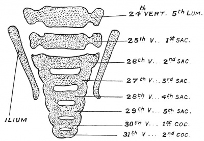
The 25th vertebra in 95 % of people forms the 1st sacral ; in 1 % the 24th, and 3 % the 26th. These percentages are drawn from the observations of Paterson, Rosenberg, and others who have made researches on this subject. The vertebral formula is not fixed. Rosenberg's investigations showed (Fig. 49) that it is the 26th vertebra that forms the first of the sacral series in the early embryo ; later the 25th throws out great lateral masses, and thus forms a connection with the ilia. Bardeen has not been able to confirm Rosenberg's observations ; he found that the vertebra which was to form the first sacral — whether it was the 24:th, 25th or 26th in the vertebral series — took on a predominance at its earliest appearance. In the lower primates (monkeys) the 27th forms the 1st sacral ; with the evolution of man the 26th, then the 25th underwent sacral modifications, the trunk being correspondingly shortened. The lumbar region of the human spine elongates much more rapidly after birth than either the cervical or dorsal region, in order to form an elongated flexible pillar for the support of the upper part of the body. In the anthropoid apes the lumbar region is relatively short as in the child at birth. It will be seen that the number of lumbar vertebrae in man is not definitely fixed. The anterior point of attachment of the ilium fluctuates from the 24th to the 26th vertebra. With the sacral transformation of the 25th and 26th (lumbar) vertebrae, there is a corresponding movement forwards of the sacral plexus.
Fig. 49. A Section of the Lumbo-sacral Region of the Spine in a Foetus at the end of the 2nd month, showing the 26th vertebra forming the 1st Sacral. (After Rosenberg.)
II. Sacro-coccygeal
The 30th vertebra forms the 1st coccygeal ; not uncommonly this vertebra is sacral in type and forms part of the sacrum. On the anterior or pelvic aspect of the 1st coccygeal vertebra a rudiment of the haemal arch is usually to be found during foetal life. The haemal arches are well developed on the proximal caudal vertebrae of tailed monkeys, and represent developments from the hypochordal or intercentral element of a vertebra. Variations at the distal end of the coccyx are dealt with later (p. 65).
III. Dorso-Iumbar region
Tins region is also liable to variation ; the 20th vertebra instead of forming the 1st lumbar, may simulate the |ast dorsal in the type of its articular processes, and may bear ribs, probably a reversion to an ancestral condition, or, on the other hand, the 12th dorsal vertebra (19th) may not carry ribs. About 2 % of bodies show the latter kind of variation — a reduction of the costal series, and about 6 to 8 % the former kind, in which the costal series is increased (see also Chap. XIX.).
IV. Dorso-cervical
The 7th vertebra may carry ribs ; rarely the 8th vertebra has no ribs attached to it and is cervical in type.
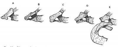
In Fig. 50 is represented the condition of the seventh cervical vertebra, as seen in 72 human skeletons. In the foetus, the costal element is always apparent ; in the adult it may vanish or fuse with the transverse process. In about 1 % of individuals it assumes the development shown in Fig. 50, E ; it may, in occasional cases, assume all the characters of a first dorsal rib, with its anterior end implanted on the presternum. Recently Prof. Wingate Todd has published a series of observations, which are confirmed by the statements made here. A cervical rib may fuse with the costal element of the first dorsal vertebra, thus giving rise to a bicipital rib (Dr. Wood Jones). The lower trunk of the bracbial plexus crosses a cervical rib, and hence in such cases symptoms of nerve-pressure may arise.
Fig. 50. Diagram showing the variation in the development of the costal element of the seventh Cervical Vertebra in 72 skeletons. In A and B the costal element is partly fused with the transverse process ; in C, D and E it remains free.
V. Cervico-occipital Region
he occipital or posterior part of the skull represents three united vertebrae.[2] Very rarely the last of these may partly assume a vertebral form, but it is by no means rare to see the atlas or first cervical vertebra partly fused with the occipital bone, representing a tendency to add a fourth vertebra to the occipital series.
The Notochord
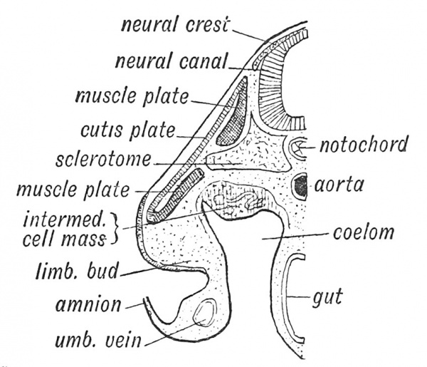
In its primitive form, this predecessor of the vertebral column is well seen during the larval stage of certain fishes (Fig. 40, A). Its manner of origin in the human embryo has been mentioned already (p. 36). The notochord when first laid down under the neural plate of the embryo is hollow — the hinder end of the canal opening at first at the neurenteric canal. Later the notochordal tube is produced in the primitive streak and later still at the growing point of the tail (Fig. 63). Afterwards the notochord becomes a solid rod composed of cells of a peculiar type. A sheath is formed round it by cells of the paraxial mesoblast (Fig. 51), which grow inwards and surround it. These cells form the sclerotome and spring from the inner parts of the primitive segments or somite into which the paraxial mesoblast is divided (p. 51). At the same time the cells of the sclerotome also grow up and gradually surround the neural tube. From these cells which grow inwards and surround the notochord and neural canal, the membranous basis of the spinal column is formed and also the basi-occipital and part of the basi-sphenoid bones of the skull (Fig. 52).
Fig. 51. A Schematic Section of an Embryo to show the sclerotome, muscle plate and skin plate which arise from each segment of the paraxial mesoblast. (Compare with Fig. 65, p. 67.)
What becomes of the Notochord
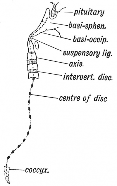
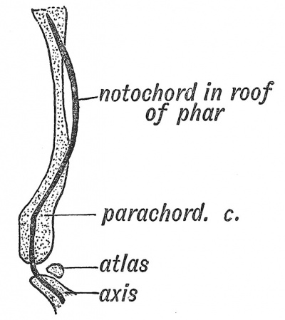
In the second month of foetal life the notochord begins to disappear[3] ; the bodies of the vertebrae and parachordal cartilages form round its sheath and constrict it. The parachordal cartilages are transformed into the basi-occipital and part of the basi-sphenoid — the basal part of the skull — behind the pituitary fossa. The notochord disappears in the basilar cartilage of the skull. Eternod, however, found the anterior part of the notochord on the dorsal wall of the pharynx in the human embryo ; Robinson has shown that in man the parachordal cartilages are developed in part on its dorsal aspect (Fig. 53). The odontoid process represents the body of the atlas, and the suspensory ligament the disc between the occipital bone and atlas. A remnant of the notochord is enclosed in the suspensory ligament. The centrum or body of each vertebra is formed round the notochord (Fig. 56, F), but only between the centra, where the intervertebral discs are formed, does this primitive structure persist. In the discs the notochord swells out and forms a considerable part of the central mucoid core which each disc contains.
Fig. 52. Where remnants of the Notochord may occur in the Adult.
Primitive Segments or Somites
Somites, or protovertebrae as they were formerly named, are not the forerunners of the vertebrae ; they are the primitive segments into which the mass of mesoderm at each side of the neural canal and notochord divides (Fig. 51, also Fig. 19), The process of division or segmentation begins at the occipital region towards the end of the third week, and spreads backwards until 35 or more body segments or somites are isolated. Each segment thus separated forms its own muscles (from its muscle plate or myotome), has its own nerve (spinal nerve), its own artery (intercostal), its own cutis plate or dermatome, and the basis for its skeletal tissue (sclerotome) (Fig. 51). The intersegmental septum separates one somite from another. Ribs, transverse and spinous processes,, are formed in the intersegmental septa. Hence an intercostal space with its muscles, vessels, and nerves, with the corresponding intervertebral structures, represents a differentiated somite. In the ventral aspect of the neck and loins, some of the intersegmental septa disappear.
Fig. 53. The relationship of the Notochord to the basilar or parachordal cartilage of the human embryo. (Arthur Robinson.)
Morphological Parts of a Vertebra
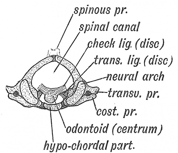
The constituent parts of a vertebra, although much modified, may be best recognized in the altas (see Fig. 54). These parts are (1) the centrum, which forms the odontoid process ; (2) the right and (3) the left half of the neural arch ; (4) the hypochordal part, which forms the anterior arch or bow. Besides the four chief elements there are three secondary processes or levers, all of which spring from the neural arch. These are (a) spinous, (6) transverse, (c) costal processes. In the dorsal region the costal processes become separated from the neural arches by articulations ; in other vertebrae they retain their continuity with the arch.
Fig. 54. The Morphological Parts of the first Cervical Vertebra.
Development of a Typical Vertebra — the 6th Dorsal
1. Membranous Stage (5th and 6th weeks)
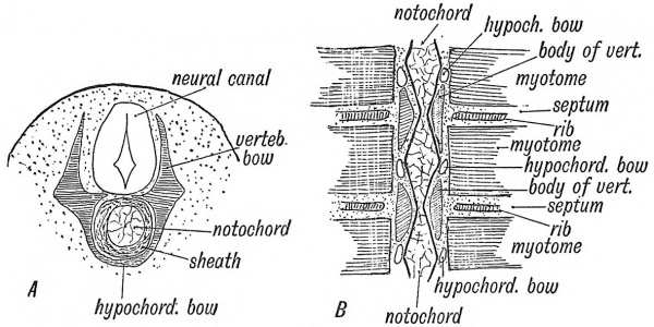
The vertebra[4] then consists of 1st a centrum surrounding the notochord, formed from its sheath (Fig. 55, A), and 2nd a horse-shoe shaped vertebral bow (Fig. 55, A and B). The bow consists of the right and left limbs which become corresponding parts of the neural vertebral arch and the hypochordal bow which unites the neural arch limbs ventral to the centrum.
Fig. 55. The development of the Membranous Basis of a Vertebra. A. In transverse section. B. In horizontal section showing the relation of the vertebra to the Primitive Segments. The section is viewed from the dorsal aspect.
2. Cartilaginous Stage
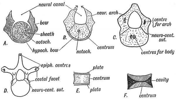
(Fig. 56) commences in the 6th week when the embryo is 9 to 10 mm. in length. The fibrous basis of the whole vertebra is transformed into cartilage. In each lateral half of the cellular basis of a vertebra three centres of chondrification appear — one for the neural arch, one for the costal process, and one for each half of the centrum, but those of the centrum soon fuse. In the process of chondrification the cells derived from the sclerotome are directly transformed into cartilage cells. In the atlas the hypochordal part of the bow becomes cartilaginous and subsequently ossified ; in all the other vertebrae, excepting the cervical segments just behind the atlas (Fig. 58), this element never passes beyond the membranous stage of development. It should be noticed (Fig. 55, B) that the vertebral bodies are formed round the notochord, opposite each intersegmental septum. Hence each centrum must be regarded as the product of two somites. The intervertebral disc is situated opposite the middle of a segment (Ebner). The lateral limbs of the cartilaginous bow meet behind (dorsal to) the neural canal in the 4th month, thus completing the neural arch. At the site of a spina bifida (see p. 83) this union fails.
Fig. 56. Showing the Stages in the Development of a Vertebra. A. In the Membranous Stage. B. In the Cartilaginous Stage. C. The appearance of Ossiflc Points. D. The appearance of Secondary Ossiflc Centres. E. The epiphyseal plates of the centra. F. Section of an amphicoelous vertebra.
3. Bony Stage
The centrum and neural arch elements of the cartilaginous vertebra fuse and give rise to the condition shown in Fig. 56, C. In the 7th week two centres of ossification appear in the centrum, but quickly fuse ; one appears in each limb of the neural arch (8th week) ; at birth the ossific centres of the centrum and neural arch have met. The central and neural ossifications meet at the neuro-central suture, and unite at the 4th or 5th year, the body being formed by^(l) the centrum, (2) basal parts of the neural arch (Figs. 54, 56). The neural ossifications fuse behind (where the spinous process is produced) in the 1st year. The spinous and transverse processes are formed by outgrowths of cartilage into the septa between the somites or primitive segments, where they serve as levers on which the spinal musculature acts. The ribs are also formed by outgrowths from the vertebrae. In the cervical, lumbar and sacral regions they fuse with the transverse processes, but in the dorsal region they remain as separate elements. In typical ribs the head corresponds to the intervertebral disc, because according to Gadow the rib was originally evolved from an intervertebral element — the intercentral or hypochordal. In atypical ribs — ^the 1st, 11th and 12th — the head of the rib articulates only with the vertebra behind its own disc. Epij)hyseal centres for the ossification of the transverse and spinous processes appear about puberty.
The Bodies of Mammalian Vertebrae are peculiar (1) in the development of an upper and lower epiphyseal plate ; (2) in that no trace of the notochord remains within them. In Fishes, as in the early human or mammalian foetus, the bodies are hour-glass shaped (amjDhicoelous, Fig. 56, F) ; in Amphibians they may retain a concavity in front (procoelous) or behind (opisthocoelous), but in mammals both ends are filled up.
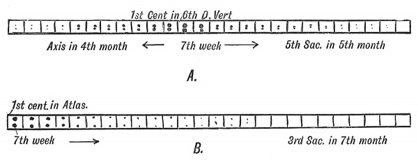
Fig. 57. The Order in which the Centres of Ossification appear in the Bodies (A) and in the Neural Arches (B) of the Spinal Column.
It will be observed (Fig. 57, B) that the centres of ossification[5] for the neural arches appear first in the anterior end of the spine (1st cervical), the date becoming later the more posterior the vertebra. In the 1st sacral they appear about the 4th month ; in the 2nd sacral, in the 5th month or later ; in the 3rd they may not appear. In the 4th and 5th sacral and 1st coccygeal vertebrae only vestiges of the neural arches are formed. These vertebrae retain the early foetal type shown in Fig. 56, B. In the remaining coccygeal vertebrae only the centres for the bodies appear. The centres for ossification of the bodies of the vertebrae appear first in the mid-dorsal region (6th dorsal). From that point they spread forwards and backwards, the centres for the odontoid process appearing at the 4th month, and that for the 5th sacral at the 5th month, while the coccygeal do not appear until about birth.
The Atlas and Axis
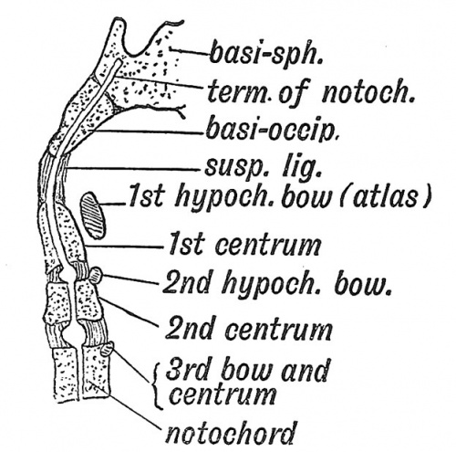
The atlas represents the completed bow of the 1st cervical vertebra (Fig. 54). The body of the vertebra fuses with the body of the 2nd, and forms the odontoid process. A remnant of the disc between the 1st and 2nd vertebrae can sometimes be seen when the odontoid is split open. The suspensory and check liagments are the representatives of the disc between the last occipital segment and the 1st cervical (Figs. 54 and 58). A nodule in the suspensory ligament may represent an occipital centrum (see p. 144).
Fig. 58. A Diagrammatic Section of the Foetal Axis, Atlas, and Basi-occipital.
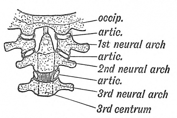
Occipito-atlanto-axial Articulations
In the intervertebral discs of the cervical region there is at each side, between the lateral lips of the vertebral bodies, a small articular cavity[6] (Fig. 59). It is situated between the part of the body formed by the neural arches and lies in front of (ventral to) the issuing spinal nerves. Between the axis and atlas this articulation is greatly enlarged. Here the rotatory movements of the atlas on the axis take place. The atlanto -occipital joint, which separates the atlas and the last occipital segment, is of the same nature. The atlas has neither the upper nor the lower articular processes of the other vertebrae. Hence the 1st and 2nd cervical nerves appear to issue behind the articular processes. At one time the single median occipital condyle seen in birds and reptiles was regarded as very different in nature from the double condyles of mammals. Eecently Symington has sliown that in the lowest mammals (mouotremes), the occijHtal condyles are fused in the middle line, and that all foetal mammals also show this condition. The articular facets on the upper surface of the atlas are also continuous over the hypochordal element. In the human skull a remnant of this median fusion of the condyles is frequently seen on the anterior margin of the foramen magnum ; it is named the third or median occipital condyle.
Fig. 59. The nature of the Atlanto-axio-occipital Articulations.
The Ribs
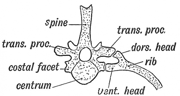
The Ribs are developed as outgrowths of the membranous vertebrae into the septa between the primitive segments of the thoracic region of the embryonic body. In lower vertebrates (birds, reptiles, etc.) each rib has two heads, a dorsal and ventral (Fig. 60) . The tuberosity of the human rib represents the dorsal head ; the ventral head is well developed in man, as in mammals generally. The rib articulates with the neural arches only (Fig. 56, B). The conjugal ligament is made up of fibres which cross in the posterior aspect of the intervertebral disc and unite the heads of the corresponding right and left rib. The conjugal ligament which is strong in some animals is weak in man (Bland-Sutton). The transverse ligament of the atlas may belong to the conjugal series.
Fig. 60. The Bicipital Rib of a Lower Vertebrate (crocodile).
Vestigial Ribs
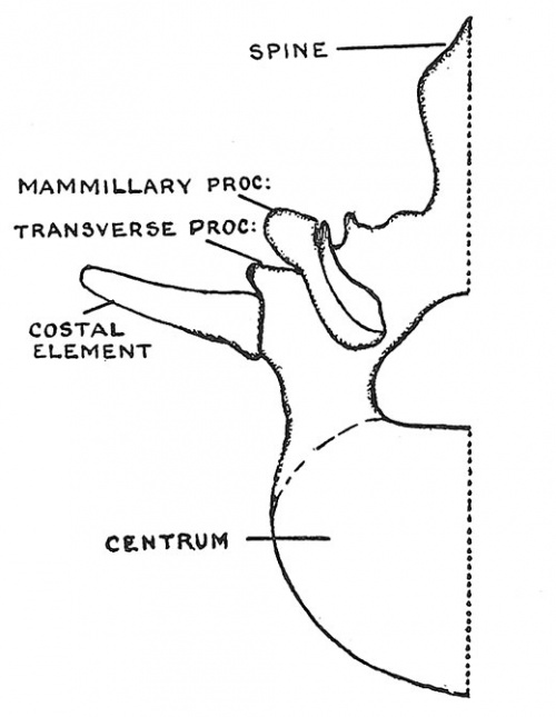
Although the ribs are only fully developed in the dorsal region, yet a representative — a costal element — is present in every vertebra. In the cervical vertebrae (Fig. 54) the anterior part of the transverse processes represents a costal process, but only in the 6th (sometimes) and 7th is the costal process formed by a separate centre of ossification. The costal process of the 7th, usually represented by a mere vestige, may develop into a rudimentary or even a fully formed rib which reaches the sternum. In the lumbar vertebrae only the first shows a separate centre for the formation of the costal process ; it fuses with the transverse process in the later months of foetal life ; in the other lumbar vertebrae the tips or perhaps the whole of the transverse processes represent costal processes. The 12th dorsal rib varies greatly in size ; it may be six or ten inches long or reduced to a mere vestige. In quite 40 % of women the 12th rib cannot be palpated because it does not project beyond the outer border of the erector spinae.
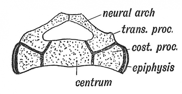
In the 1st, 2nd and 3rd sacral vertebrae the costal processes are large and have their own centres of ossification.[7] Their cartilaginous bases fuse early to form the greater part of ttie lateral masses of the sacrum. The part of the lateral mass formed by the costal processes is shown in Fig. 62. The costal processes are absent in the 4th and 5th sacral and in all the coccygeal vertebrae. The two lateral epiphyseal plates on each side of the sacrum are new and independent formations.
The Accessory Processes are found in the lumbar and lowest two dorsal vertebrae. They are developed at the base of the transverse processes and are for the attachment of slips of the longissimus dorsi. The mammillary processes are developed on the articular processes of the lower two or three dorsal and all the lumbar vertebrae. They give attachment to tendons of origin of the multifidus spinae. In a paper {Journ. Anat. and Physiol. 1912, vol. 47, p. 118) by Dr. Wood Jones, it is pointed out that these two muscular processes, the mammillary and accessory, are fused together in the dorsal region, but in the lumbar region they are separated by a groove containing the inner branch of the posterior division of the corresponding spinal nerve.
Fig. 61. Half of a first Lumbar Vertebra showing a separate costal element.
Fig. 62. A Section to show the Nature of the Elements composing the Sacrum.
The Transverse and Spinous Processes
The Transverse and Spinous Processes grow out from the vertebral bow (Fig. 56, A) into the septa between the primitive segments. Each transverse process is pierced, while still in the fibrous condition, by a branch of the corresponding segmental (intercostal) artery. In only the cervical region do those perforating arteries and their foramina persist. In that region the perforating arteries anastomose, and out of the chain thus formed is developed the vertebral artery. Thus the foramina for the vertebral artery are formed independently of the costal element in each cervical transverse process. The spines are absent on the 1st cervical, 4th and 5th sacral and coccygeal vertebrae. They are slightly developed and united by ossification of the interspinous ligament in the 2nd and 3rd sacral vertebrae. The 2nd, 3rd, 4th, 5th, and 6th cervical spines are bifid in Europeans ; but in lower races, as in anthropoids, the 5th and 6th spines are usually undivided.
Caudal or Coccygeal Vertebrae
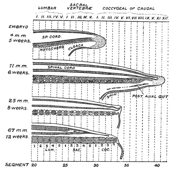
At the end of the 6th week, the body of the embryo being then 11 mm. in length, the human tail reaches its maximum growth — projecting as a conical process fully 1 mm. in length and equal to about one-tenth of the long diameter of the embryonic body. En the adult body the 30th vertebra is usually the first of the coccygeal series. In the fifth week the growing caudal point, at which neural canal, notochord, sclerotomes, and cloaca are all being extended in a backward direction, has reached and produced the 30th segment (Fig. 63) ; at the 6th week, ten or twelve caudal segments have been laid down. Thereafter retrogression sets in ; by the end of the 8th week (Fig. 63) only the caudal tip j)rojects and the coccygeal vertebrae have been reduced to 4 or 5, while, by the 13th week, a depression or pit marks the site where the tip disappeared. The coccygeal part of the neural canal is atrophied and the distal part of the whole cord is retracting in a cranial direction.[8]
Fig. 63. A series of four figures showing the conlition of the liuman caudal or coccygeal region at the stages indicated on the drawings (after Kunitomo).
- ↑ For literature on variations of vertebrae : Bardeleben, Ergebnisse der Anat. 1905, vol. 15, p. 119 ; 1906, vol. 16, p. 141 ; 1908, vol. 18. p. 71. A. Fischel, Anat.\Hefte, 1906, vol. 31, p. 459. E. Rosenberg, Morph. Jahrbuch, 1907, vol. 36, p. 609. F. Wood Jones, Journ. Anat. and Physiol. 1910, vol. 44, p. 377 (Influence of Nerve -plexuses in determining Development of Costal Processes). C. R. Bardeen, A^ner. Journ. Anat. 1905, vol. 4, p. 163 (Development of Vertebrae). E. Barclay Smith, Journ. Anat. and Physiol. 1911, vol. 45, p. 144. T. Manners Smith, Journ. Anat. and Physiol. 1909, vol. 43, p. 146. A. F. Le Double, Bull, et Mem. Soc. d'Anthrop. 1911, Ser. 6, vol. 2, p. 413 (Lumbar Ribs) ; p. 428 (Cervical Ribs). F. Wood Jones, Journ. Anat. and Physiol. 1911, vol. 45, p. 249 (Cervical Ribs). T. W. Todd, Journ. Anat. and Physiol. 1912, vol. 46, p. 244 (Cervical Ribs). J. C. Brash (Anomalous Spines), Journ. Anat. 1915, vol. 49, p. 243. M. F. Lucas-Keen, Journ. Anat. 1915, vol. 49, p. 336.
- ↑ For reports of cases of fusion of atlas : Schumacher, Anat. Anz. 1907, vol. 31, p. 145 (Homologies of Occipital Bone) ; K. Weigner, Anat. Hefte, 1911, vol. 45, p. 81 (Assimilation of Atlas) : Glaesmer, Anat. Anz. 1910, vol. 36, p. 129 ; Le Double, Bull, et Mem. Anat. 1912, p. 20 ; G. Elliot Smith, Brit. Med, Jgurn, 1908, 2, p. 594 . R. J. Gladstone, Journ. Anat. 1915, vol, 49, p. 190,
- ↑ Papers on notochord: A. Bruni, Anat. Hefte, 1912, vol. 45, p. 307 (Involution of Notochord) ; A. Linck, Anat. Hefte, 1911, vol. 42, p. 607 (Dev. of Notochord) ; L, W. Williams, Amer, Journ. Anat. 1908, vol. 8, p. 251
- ↑ For an account of the differentiation and development of vertebrae : C. R. Bardeen, Amer. Journ. Anat.,\Q05, vol. 4, p. 163 (Thoracic Vertebrae) ; also p. 265 ; 1908, vol. 8, p. 181 (Cervical and Occipital Regions).
- ↑ See F. P. Mall, Amer. Journ. Anat. 1906, vol. 5, p. 433 (Centres of Ossification before end of 2nd month); E. Fawcett, Journ. Anat. and Physiol. 1911, vol. 45, p. 172 (Costal Epiphyses). Prof. F, Dixon, Journ. A^iat. 1921, vol. 55, p. 38.
- ↑ 0. Jaekel, Anat. Anz. 1912, vol. 40, p. 609 (Morphology of Atlas).
- ↑ References to papers on sacrum : E. Fawcett, A7iat. Anz. 1907, vol. 30, p. 414 (Sacral Costal Epiphyses) ; Otto Petersen, Anat. Anz. 1905, vol. 26, p. 521 ; D. E. Derry, Journ. Anat. and Physiol. 1911, vol. 45, p. 202 (Sacral Accessory Articulations) L. Bolk, Anat. Anz. 1912, vol. 41, p. 54.
- ↑ See Prof. G. L. Streeter, Amer. Journ. Ayiat. 1919, vol. 25, p. 1 ; Dr. Kanae Kunitomo, Contrib. to Embryology, 1918, vol. 8, p. 161.
| Historic Disclaimer - information about historic embryology pages |
|---|
| Pages where the terms "Historic" (textbooks, papers, people, recommendations) appear on this site, and sections within pages where this disclaimer appears, indicate that the content and scientific understanding are specific to the time of publication. This means that while some scientific descriptions are still accurate, the terminology and interpretation of the developmental mechanisms reflect the understanding at the time of original publication and those of the preceding periods, these terms, interpretations and recommendations may not reflect our current scientific understanding. (More? Embryology History | Historic Embryology Papers) |
Human Embryology and Morphology: 1 Early Ovum and Embryo | 2 Connection between Foetus and Uterus | 3 Primitive Streak Notochord and Somites | 4 Age Changes | 5 Spinal Column and Back | 6 Body Segmentation | 7 Spinal Cord | 8 Mid- and Hind-Brains | 9 Fore-Brain | 10 Fore-Brain Cerebral Vesicles | 11 Cranium | 12 Face | 13 Teeth and Mastication | 14 Nasal and Olfactory | 15 Sense OF Sight | 16 Hearing | 17 Pharynx and Neck | 18 Tongue, Thyroid and Pharynx | 19 Organs of Digestion | 20 Circulatory System | 21 Circulatory System (continued) | 22 Respiratory System | 23 Urogenital System | 24 Urogenital System (Continued) | 25 Body Wall and Pelvic Floor | 26 Limb Buds | 27 Limbs | 28 Skin and Appendages | Figures

