Human Embryology and Morphology 4
Keith, A. Human Embryology And Morphology (1921) Longmans, Green & Co.:New York.
Human Embryology and Morphology: 1 Early Ovum and Embryo | 2 Connection between Foetus and Uterus | 3 Primitive Streak Notochord and Somites | 4 Age Changes | 5 Spinal Column and Back | 6 Body Segmentation | 7 Spinal Cord | 8 Mid- and Hind-Brains | 9 Fore-Brain | 10 Fore-Brain Cerebral Vesicles | 11 Cranium | 12 Face | 13 Teeth and Mastication | 14 Nasal and Olfactory | 15 Sense OF Sight | 16 Hearing | 17 Pharynx and Neck | 18 Tongue, Thyroid and Pharynx | 19 Organs of Digestion | 20 Circulatory System | 21 Circulatory System (continued) | 22 Respiratory System | 23 Urogenital System | 24 Urogenital System (Continued) | 25 Body Wall and Pelvic Floor | 26 Limb Buds | 27 Limbs | 28 Skin and Appendages | Figures
| Historic Disclaimer - information about historic embryology pages |
|---|
| Pages where the terms "Historic" (textbooks, papers, people, recommendations) appear on this site, and sections within pages where this disclaimer appears, indicate that the content and scientific understanding are specific to the time of publication. This means that while some scientific descriptions are still accurate, the terminology and interpretation of the developmental mechanisms reflect the understanding at the time of original publication and those of the preceding periods, these terms, interpretations and recommendations may not reflect our current scientific understanding. (More? Embryology History | Historic Embryology Papers) |
Chapter IV. The Age Changes in the Embryo and Foetus
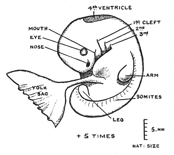
In Chapter I., having followed the developmental changes in the human embryo during the first five weeks, when it had reached a crown-rump length of 5 mm. {1/5 in.) and the condition of parts shown in Fig. 42, we had to break away in Chapters II. and III. to note the manner in which it effected a lodgment in the uterus and to examine certain processes which give rise to fundamental parts of the embryonic body. In this chapter we return to trace the further history of the embryo, to watch it becoming transformed into a foetus and to register the subsequent changes during the nine months it spends in its mother's womb.
Fig. 42. Outline of a Human Embryo 5 mm. in length, and in the 5th week of development. (Reconstructed by Professor Keibel and Dr. Elze.)
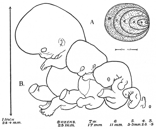
In recent years our knowledge concerning the rate at which the human embryo grows and the stages through which it passes week by week has become more accurate. This is largely due to the work accomplished by the late Prof. Mall[1] who collected facts relating to all cases where the age of an embryo had been ascertained and by tabulating his data was able to estimate the size and stage of development reached by an average human embryo week after week. His main results, so far as concern the first two months, are set out in Fig. 43, taken from an article written by his distinguished pupil — Prof. Herbert Evans. Six stages of development are represented : at the end 3rd, 4th, 5th, 6th, 7th and 8th weeks. Under each embryo is given the mean length it should reach at a certain date, but it has to be remembered that the rate of growth will vary in embryos and foetuses just as in children, and that some will be precocious while others will be backward. The measurements relate to fresh specimens, for when embryos are preserved and prepared for microscopic examination they shrink in size. It is convenient to regard 3 mm. as measured from the crown to the rump of the embryo as marking the end of the fourth week, and 5 mm. as an index of the end of the fifth week of development. In the 6th, 7th, and 8th weeks the embryo adds almost one millimetre to its length daily, being about 25 mm. (1 inch) at the end of the 8th week. Hence we may readily estimate the age of an embryo or foetus under 25 mm. in length, by regarding the first 5 mm. as representing 35 days' growth, and adding a day for every additional millimetre of its length. For example, the age in days of an embryo measuring 15 mm. in length would be estimated thus : 5 mm. = 35 days + 10 for the additional 10 mm. = 45 days. In the 9th, 10th, 11th, 12th, 13th and 14th weeks — up to the end of the 3rd month — when the crown-rump length amounts to 100 mm. (4 in.)^ — the daily rate of growth is approximately 1"5 mm.
Fig. 43. Series of six drawings illustrating the stages of growth from the end of the 3rd week to the end of the 8th. In a corner of the figure is a diagram to illustrate the rate of growth of the chorionic vesicle at corresponding dates. (Prof. H. M. Evans.)
External Changes in the 6th week
As may be seen by comparing Figs. 42 and 44, the 6th week[2] constitutes a period of rapid transformation.
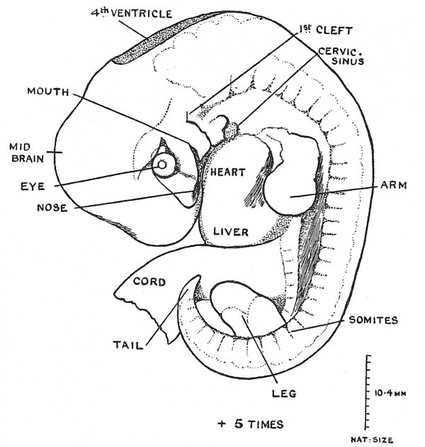
Not only does the length of the embryo increase from 5 mm. to 11 mm. but there are very definite changes in the form of its external parts. At the end of the 5th week the gill-arch system of the primitive pharynx is at its height, four arches being distinguishable ; in the 6th week the 3rd and ith arches sink into a pit in the neck — the cervical sinus — (Fig. 44), while the 2nd or hyoid arch grows backwards over the pit and thus hides the hinder arches. Here we are witnessing the closing in or operculation of the branchial arches — as it takes place in gill-bearing vertebrates. Even at the close of the 6th week the face is represented merely by a forehead filled out by the relatively small f orebrain vesicle ; behind and under the forehead are seen the nasal, maxillary and mandibular processes which will give rise to the face proper. All of these elements have made headway during the 6th week (Figs. 42, 43, 44). The head region even in the 6th week is still tubular in form ; the mid and hind brains form the greater part of the central nervous system, for the cerebral vesicles have as yet only begun to grow out from the fore brain. The limb buds, which in the 5th week were still undemarcated into segments, now show their three primitive parts — upper arm and thigh, forearm and leg, and a platelike hand and foot. In point of differentiation the forelimb is always in advance of the hind. At the close of the 6th week the first appearance of jvebbed digits can be detected, and at the same time, when the length of the embryo is about 11 mm. the tail reaches its maximum development (Fig. 44) ; in the 7th week retrogression has already set in. The umbilical cord becomes lengthened and more clearly differentiated in the 6th week ; between the attachment of the cord to the ventral wall of the embryo and the gill-formation of the primitive pharynx is seen the bulging eminence of the heart (Fig. 42) ; below the heart eminence, as may be seen in Fig. 44, there appears in the 6th week a second eminence, that caused by the developing liver.
Fig. 44. Outline of a Human Embryo 10.4 mm. long and in the 6th week of development. (After Broman.) (Mag. x 5.)
Embryo-Foetus
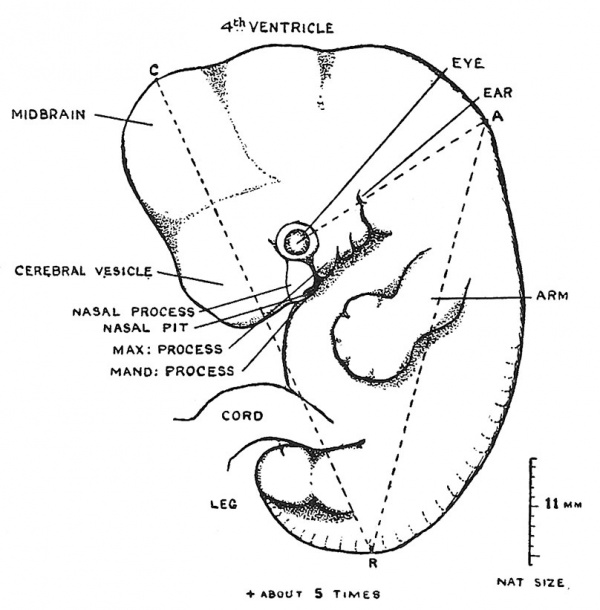
During the 7th week the embryo becomes a foetus — the transformation being well shown in Fig. 43. In its crown-rump length the embryo expands from 11 to 17 mm., but the characteristic changes are seen in the face, head and limbs. An early stage of the 7th week is shown in Fig. 45 ; the basal parts of the face are being laid down. Under the eye are seen the nasal processes carrying the open nasal cavities backwards into the region of the mouth, while growing forwards beneath the eye are to be observed the maxillary processes which will provide the bases of the upper jaw. Still further back in the pharynx ( Fig. 45) are seen two comparatively small processes — the mandibular (1st arch) and hyoid (2nd arch). Behind the hyoid arch there is a depression marking the cervical sinus. By the end of the 7th week Fig. 43) the nasal, maxillary and mandibular processes have united to form a relatively small face ; at the upper end of the postmandibular cleft has appeared the rudiment of an ear. The changes in the head itself are also apparent ; at the end of the 7th week the cylindrical cranial form is being replaced by one more distinctly globular ; the forehead in particular has become enlarged. These changes are due to the rapid expansion of the cerebral vesicles during the 7th week. The changes in the limbs are also very evident ; they are now folded on the belly-wall, palm towards palm and sole towards sole ; the digits are demarcated. The tail is disappearing. The head is no longer bent forwards with the forehead touching the root of the umbilical cord, but is lifted up, for the embryonic flexure of the cervical region is being undone and a narrowing of the post-cranial region to form a neck becomes apparent. The heart is now completely divided into right and left chambers and the growth of the neck is lifting the pharyngeal region away from the heart. With these changes in the facial region, in the head, neck, limbs and heart the embryo of the 6th week becomes the foetus of the 7th. One other very important event also characterises this stage of transformation : the cellular blastema of the skeleton begins to change into cartilage and into bone. It also becomes possible to distinguish the ovary from the testicle.
Fig. 45. Outline of an embryo, although only 11 mm. long, yet showing changes characteristic of an early stage of the 7th week. (After Broman.) The line C-R indicates the manner in which the crown-rump diameter is measured ; the line A-R shows the neck-rump length, A being found by drawing a line backward through the rudiments of the eye and ear.
Changes in the 8th week
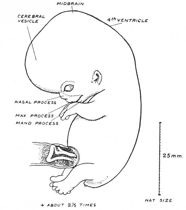
At the end of the 8th week the crownrump diameter measures about 25 mm. (1 inch). The changes of this week are a continuation of those we have just described (Fig. 46) ; the nasal and maxillary processes have fused to form the upper face ; the upper lip is completed, but the palatal processes have not yet separated the buccal from the nasal cavities. The cerebral vesicles are expanding rapidly backwards ; the neck is being differentiated and the limbs are making progress. The rudiment of the external genital organs is apparent, but as yet gives no clue to sex. The intestinal loop lies within the root of the umbilical cord. Henceforward, until the end of gestation, the chief changes are those of growth.[3]
Fig. 46. Outline of a Foetus 22 mm. long, and at the end of the 2nd month of development. (After Broman.)
The Full-Time Foetus
We speak of the period of human gestation — the time spent by the human young in the uterus of the mother, preparatory to an independent existence — as being one of 9 months, and if by a month we mean 30 days — 270 days in all — we are as near the truth as our present evidence will take us. Medical men can seldom discover the exact date of conception and hence to get a fixed point for a reckoning they begin their estimate counting from the first day of the mother's last menstrual period, and taking this day as a fixed point, count that parturition will take place 280 days hence. Observations made on cases where the date of impregnation may be inferred show that the actual mean period of gestation is 270 days. The 270th day is the bull's-eye at which Nature aims, but even the best of marksmen make " inner s " and " outers," and it is so in all of Nature's shootings. She is ever subject to the law of chance ; hence in all developmental and growth manifestation we meet with variation round a mean.
By the 270th day the foetus has attained a weight of about 7 lbs. and a height, if we measure from crown to rump (sitting height), of 336 mm., but if we include the lower extremities (standing height) the measurement is 500 mm. (nearly 20 inches). It sometimes happens that birth takes place at the end of the 7th month when the foetus weighs between 4-5 lbs. and in its standing height measures 400 mm. or less, its sitting height being then about 265 mm. In such premature children, who have always a defective heat-regulating mechanism, it will be observed that the tips of the nails just reach the ends of the nail beds, whereas in the full-time child the nail edges are free and projecting. The full-time child has also an outcrop of hair on the head ; lanugo — foetal hair — can be detected on various parts of the body. The hair tips which break on the surface of the skin about the end of the 4th month may be plentiful on the scalp at the end of the 7th, but the skin is then of bright lobster-red, the subcutaneous tissue is less stored with fat and the sebaceous covering, known as the vernix caseosa, forms a thin and unequal coating.
Table of Growth
It is impossible for anyone to remember the dimensions reached during the various stages of foetal development and growth, but it is often convenient to have a table of measurements for reference. The one given here was prepared by the late Prof. Mall[4]:
| Crown-Rump Length (mm) | Standing Height | Age in Weeks | Age in Days |
|---|---|---|---|
| 0.55 | — | 3 | 21 |
| 2-5 | — | 4 | 28 |
| 5-5 | — | 5 | 35 |
| 11 | — | 6 | 42 |
| 17 | — | 7 | 49 |
| 25 | — | 8 | 56 |
| 32 | — | 9 | 63 |
| 43 | — | 10 | 70 |
| 53 | — | 11 | 77 |
| 68 | — | 12 | 84 |
| 81 | — | 13 | 91 |
| 100 | 149 | 14 | 98 end of 3rcl month |
| 111 | — | 15 | 105 |
| 121 | — | 16 | 112 |
| 134 | — | 17 | 119 |
| 145 | 223 | 18 | 126 end of 4th month. |
| 157 | — | 19 | 133 |
| 167 | — | 20 | 140 |
| 180 | — | 21 | 147 |
| 192 | 295 | 22 | 154 end of 5th month |
| 202 | — | 23 | 161 |
| 210 | — | 24 | 168 |
| 220 | — | 25 | 175 |
| 230 | 331 | 26 | 182 end of 6th month. |
| 237 | — | 27 | 189 |
| 245 | — | 28 | 196 |
| 252 | — | 29 | 203 |
| 265 | 400 | 30 | 210 end of 7th month. |
| 276 | — | 31 | 217 |
| 284 | — | 32 | 224 |
| 293 | — | 33 | 231 |
| 301 | 443 | 34 | 238 end of 8th month. |
| 310 | — | 35 | 245 |
| 316 | — | 36 | 252 |
| 325 | — | 37 | 259 |
| 336 | 500 | — | 270 end of 9th month. |
- ↑ See his last paper on this subject, Amer. Jouni. of Anat. 1918, vol. 23, p. 397.
- ↑ See specimens described by J. L. Bremer, Amer. Journ. Anat. 1905, vol. 5, p. 459 ; C. Elze, Anat. Hefte, 1907, vol. 35, p. 409 ; N. W. Ingalls, Archiv. f. mikros. Anat. und Entwick. 1907, vol. 70, p. 506 ; L. Frassi, Ibid. p. 492. Dr. H. L. Bamiville gives a full description of an 8-5 mm. Embryo, Journ. Anat. 1915, vol. 49, p. 1. Dr. F. W. Thyng gives excellent figures of one measuring 17-8 mm., Amcr. Jottrn. of Anat. 1915, vol. 17, p. 51.
- ↑ For changes in 9th week see F. E. Blaisdell, Journ. Anat. 1914, vol. 48, p. 182.
- ↑ See his last paper on this subject, Amer. Jouni. of Anat. 1918, vol. 23, p. 397.
| Historic Disclaimer - information about historic embryology pages |
|---|
| Pages where the terms "Historic" (textbooks, papers, people, recommendations) appear on this site, and sections within pages where this disclaimer appears, indicate that the content and scientific understanding are specific to the time of publication. This means that while some scientific descriptions are still accurate, the terminology and interpretation of the developmental mechanisms reflect the understanding at the time of original publication and those of the preceding periods, these terms, interpretations and recommendations may not reflect our current scientific understanding. (More? Embryology History | Historic Embryology Papers) |
Human Embryology and Morphology: 1 Early Ovum and Embryo | 2 Connection between Foetus and Uterus | 3 Primitive Streak Notochord and Somites | 4 Age Changes | 5 Spinal Column and Back | 6 Body Segmentation | 7 Spinal Cord | 8 Mid- and Hind-Brains | 9 Fore-Brain | 10 Fore-Brain Cerebral Vesicles | 11 Cranium | 12 Face | 13 Teeth and Mastication | 14 Nasal and Olfactory | 15 Sense OF Sight | 16 Hearing | 17 Pharynx and Neck | 18 Tongue, Thyroid and Pharynx | 19 Organs of Digestion | 20 Circulatory System | 21 Circulatory System (continued) | 22 Respiratory System | 23 Urogenital System | 24 Urogenital System (Continued) | 25 Body Wall and Pelvic Floor | 26 Limb Buds | 27 Limbs | 28 Skin and Appendages | Figures

