File:Regions of varying neural cell types in ventral neural tube.jpg
Regions_of_varying_neural_cell_types_in_ventral_neural_tube.jpg (600 × 537 pixels, file size: 35 KB, MIME type: image/jpeg)
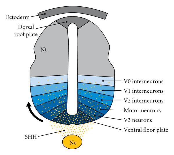
Image depicting the neural tube (nt) during embryonic development, where the regions of different inter-neurons being V0, V1, V2, and V3, along side the motor neuron (MN) region is shown. The ventral floor plate and notochord (Nc) is depicted ventrally, where the yellow dots represent sonic hedgehog (Shh) secretions by both the structures. As the Shh diffuses dorsally towards the V0 region, the concentration is diminished forming a gradient.
Copyright
Copyright © 2011 Ryan W. Y. Lee and Elaine Tierney. This is an open access article distributed under the Creative Commons Attribution License, which permits unrestricted use, distribution, and reproduction in any medium, provided the original work is properly cited.
https://creativecommons.org/licenses/by/3.0/au/deed.en
Reference
<pubmed>22937253 </pubmed>
- Note - This image was originally uploaded as part of an undergraduate science student project and may contain inaccuracies in either description or acknowledgements. Students have been advised in writing concerning the reuse of content and may accidentally have misunderstood the original terms of use. If image reuse on this non-commercial educational site infringes your existing copyright, please contact the site editor for immediate removal.
File history
Click on a date/time to view the file as it appeared at that time.
| Date/Time | Thumbnail | Dimensions | User | Comment | |
|---|---|---|---|---|---|
| current | 22:05, 27 October 2016 |  | 600 × 537 (35 KB) | Z5019880 (talk | contribs) |
You cannot overwrite this file.
File usage
The following 3 pages use this file:
