File:MRI renal agenesis .jpg
Original file (600 × 649 pixels, file size: 67 KB, MIME type: image/jpeg)
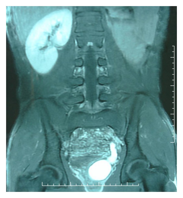
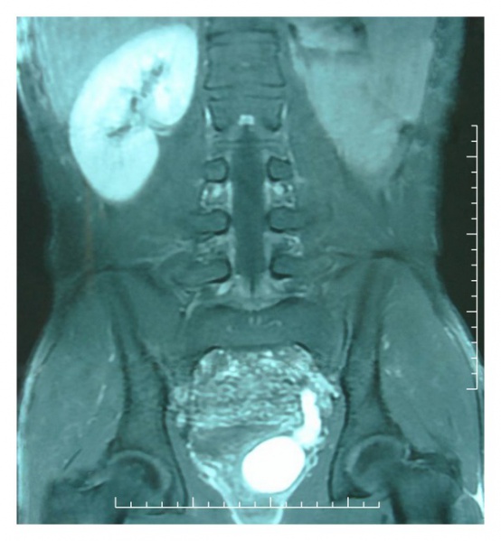
Renal agenesis
MRI showing a tube-like structure meandering along the rear wall of the bladder corresponding to the distal ureter, a cyst in the seminal vesicle area and renal agenesis.
Reference
<pubmed>22606606</pubmed>
Copyright
Copyright © 2011 Youness Ahallal et al. This is an open access article distributed under the Creative Commons Attribution License, which permits unrestricted use, distribution, and reproduction in any medium, provided the original work is properly cited.
--Mark Hill (talk) 15:10 7 November 2014 (EST) Assessment - Figure relates to project topic contains reference, copyright and student template. This abnormality needs to be put in context of fetal renal development.
- Note - This image was originally uploaded as part of an undergraduate science student project and may contain inaccuracies in either description or acknowledgements. Students have been advised in writing concerning the reuse of content and may accidentally have misunderstood the original terms of use. If image reuse on this non-commercial educational site infringes your existing copyright, please contact the site editor for immediate removal.
File history
Click on a date/time to view the file as it appeared at that time.
| Date/Time | Thumbnail | Dimensions | User | Comment | |
|---|---|---|---|---|---|
| current | 14:30, 17 September 2014 | | 600 × 649 (67 KB) | Z3465141 (talk | contribs) | =Renal agenesis= MRI imaging confirming renal agenesis ==Reference== <pubmed>22606606</pubmed> [http://www.ncbi.nlm.nih.gov/pubmed/22606606] ==Copyright== Copyright © 2011 Youness Ahallal et al. This is an open access article distributed under the... |
| 12:59, 17 September 2014 |  | 600 × 649 (67 KB) | Z3465141 (talk | contribs) |
You cannot overwrite this file.
File usage
The following 2 pages use this file: