File:Heart disection.jpg

Original file (963 × 731 pixels, file size: 209 KB, MIME type: image/jpeg)
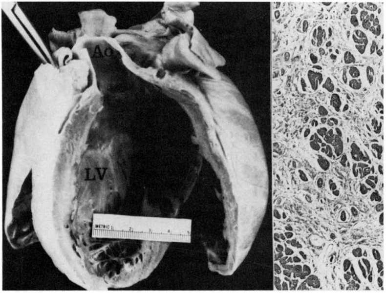
The gross specimen (left)shows the mildly dilated left ventricle(LV) with normal wall thickness; the walls were flabby. The microscopic section(right) from the left ventricular free wall shows marked connective tissue replacement. Small vessel coronary artery disease was not identified, although it was specifically sought. Ao = aorta.
Reference: J S Child, J K Perloff, P M Bach, A D Wolfe, S Perlman, R A Kark Cardiac involvement in Friedreich's ataxia: a clinical study of 75 patients. J. Am. Coll. Cardiol.: 1986, 7(6);1370-8 PMID:2940284 | J. Am. Coll. Cardiol.
Permission: Permission: None required for student projects and non-commercial use. For any commercial use of figures from JACC Imaging articles, permission is required. However if it is a student paper permission will not be needed. Please do give full attribution to the source though.
Best Regards,
Justin Byrne Managing Editor JACC Cardiovascular Imaging & JACC Cardiovascular Interventions 3655 Nobel Drive, Suite 630 San Diego, CA 92122 Tel: 858-558-3411 Fax: 858-558-3148 email: jbyrne@acc.org
- Note - This image was originally uploaded as part of a student project and may contain inaccuracies in either description or acknowledgements. Students have been advised in writing concerning the reuse of content and may accidentally have misunderstood the original terms of use. If image reuse on this non-commercial educational site infringes your existing copyright, please contact the site editor for immediate removal.
Cite this page: Hill, M.A. (2024, April 26) Embryology Heart disection.jpg. Retrieved from https://embryology.med.unsw.edu.au/embryology/index.php/File:Heart_disection.jpg
- © Dr Mark Hill 2024, UNSW Embryology ISBN: 978 0 7334 2609 4 - UNSW CRICOS Provider Code No. 00098G
File history
Click on a date/time to view the file as it appeared at that time.
| Date/Time | Thumbnail | Dimensions | User | Comment | |
|---|---|---|---|---|---|
| current | 17:59, 3 October 2011 |  | 963 × 731 (209 KB) | Z3329495 (talk | contribs) | The gross specimen (left)shows the mildly dilated left ventricle(LV) with normal wall thickness; the walls were flabby. The microscopic section(right) from the left ventricular free wall shows marked connective tissue replacement. Small vessel coronary ar |
You cannot overwrite this file.
File usage
The following page uses this file: