File:Figure Shows How the CNS is divided to supply different structures.jpg
Figure_Shows_How_the_CNS_is_divided_to_supply_different_structures.jpg (585 × 549 pixels, file size: 123 KB, MIME type: image/jpeg)
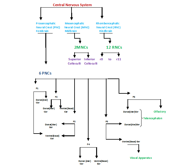
Figure Shows How the CNS is divided to supply different structures
The forebrain is made up of six prosomeres. These have designated numbers from caudal to cranial. Each prosemere are subdivided into two tiers, dorsal (alar) and ventral (basal). The alar tiers of p6 and p5 gives rise to the telencephalon while the basal tier of p5 and p6 are associated with the visual apparatus and the olfactory respectively. The midbrain is made up of two mesomeres, m1 and m2. These contain the superior and inferior colliculi respectively. The 12 rhombomeres, numbered r0 to r11 gives rise to the hindbrain. [1]
A copyright statement: "Beginning six months after publication, I z3308968 grant the public the non-exclusive right to copy, distribute, or display the Work under a Creative Commons Attribution-Noncommercial-Share Alike 3.0 Unported license, as described at http://creativecommons.org/licenses/by-nc-sa/3.0/ and http://creativecommons.org/licenses/by-nc-sa/3.0/legalcode."
File history
Click on a date/time to view the file as it appeared at that time.
| Date/Time | Thumbnail | Dimensions | User | Comment | |
|---|---|---|---|---|---|
| current | 22:59, 19 September 2011 | | 585 × 549 (123 KB) | Z3308968 (talk | contribs) | Figure Shows How the CNS is divided to supply different structures The forebrain is made up of six prosomeres. These have designated numbers from caudal to cranial. Each prosemere are subdivided into two tiers, dorsal (alar) and ventral (basal). The alar |
You cannot overwrite this file.
File usage
The following file is a duplicate of this file (more details):
The following page uses this file: