File:Expression of CD82 in human placental villi and cell lines.JPG
Expression_of_CD82_in_human_placental_villi_and_cell_lines.JPG (612 × 432 pixels, file size: 75 KB, MIME type: image/jpeg)
Description
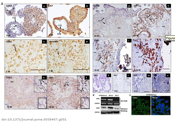
Expression of CD82 in human placental villi and cell lines.
(A) Immunostaining of CD82 in normal human placental villi in the first trimester, maternal decidua, second trimester and third trimester. (a) CD82 was strongly expressed in trophoblast columns (TC) and moderate in CTB of human placental villi during the first trimester. (c) CD82 was highly expressed in decidual cells (DC). (e) CD82 was highly expressed in the maternal decidua, not detected in EVT and very faint in anchoring villous during the early second trimester. Boxed region is enlarged on the upper panel. (g) CD82 was absent in villous and very faint in EVT during the late second trimester. (i) CD82 was moderate expressed in cytotrophoblast cells invaded into the maternal decidua and highly expressed in syncytiotrophoblast in the third trimester. (b, f, h, j) Immunohistochemical staining with anti-cytokeratin7 (CK7) as a marker of CTB, TC in the first trimester placental vill, EVT in the maternal decidua; (d) Immunohistochemical staining with anti-vimentin as a maker of decidual cells. (k, l, m, n) negative controls (NEG) on sections in which normal IgG was used in place of primary antibody. CTB: cytotrophoblast; STB: syncytiotrophoblast; TC: trophoblast column; EVT: extravillous trophoblast; MD: maternal decidua, AV: anchoring villous W: weeks of pregnancy; Bar represents 100 µm. (B) Expression of CD82 in different trophoblast cell lines determined by semiquantitative RT-PCR and Western blotting, respectively. GAPDH was used as an internal control for RT-PCR or loading control for Western blotting. HTR8/SVneo: a human invasive extravillous trophoblast cell line derived from immortalized first trimester trophoblast; B6Tert: immortalized cytotrophoblast cell line; JEG-3: human choriocarcinoma cell lines. (C) Immunofluorescence of CD82 in HTR8/SVneo cell lines. Fluorescence signals specific to CD82 antibody were visualized as green, and the nuclei were shown by DAPI staining (blue).
References
<pubmed> 22679510 </pubmed>
Citation: Zhang Q, Tan D, Luo W, Lu J, Tan Y (2012) Expression of CD82 in Human Trophoblast and Its Role in Trophoblast Invasion. PLoS ONE 7(6): e38487. doi:10.1371/journal.pone.0038487
Copyright: © 2012 Zhang et al. This is an open-access article distributed under the terms of the Creative Commons Attribution License, which permits unrestricted use, distribution, and reproduction in any medium, provided the original author and source are credited.
Expression of CD82 in human placental villi and cell lines.
http://www.plosone.org/article/info%3Adoi%2F10.1371%2Fjournal.pone.0038487
- Note - This image was originally uploaded as part of an undergraduate science student project and may contain inaccuracies in either description or acknowledgements. Students have been advised in writing concerning the reuse of content and may accidentally have misunderstood the original terms of use. If image reuse on this non-commercial educational site infringes your existing copyright, please contact the site editor for immediate removal.
File history
Click on a date/time to view the file as it appeared at that time.
| Date/Time | Thumbnail | Dimensions | User | Comment | |
|---|---|---|---|---|---|
| current | 13:45, 7 August 2012 | | 612 × 432 (75 KB) | Z3333794 (talk | contribs) | Expression of CD82 in human placental villi and cell lines. http://www.plosone.org/article/info%3Adoi%2F10.1371%2Fjournal.pone.0038487 |
You cannot overwrite this file.
File usage
The following page uses this file: