File:Electroencephalography of Angelman Syndrome.jpg
Electroencephalography_of_Angelman_Syndrome.jpg (440 × 207 pixels, file size: 33 KB, MIME type: image/jpeg)
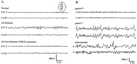
Electroencephalography of Angelman Syndrome
Electroencephalography (EEG) showing Normal and abnormal EEGs in both human and mouse.
A.
- Normal, Segment of routine EEG on a normal 10-yr-old male with no seizures.
- AS-Deletion, Background EEG of a 9.5-yr-old male, large deletion AS case.
- AS-Non-Deletion, Background EEG of a 10-yr-old male with a UBE3A gene loss-of-function mutation.
The above illustrates that seizures are most severe in AS cases with deletion of the UBE3A gene.
B.
- Background EEGs from mouse littermates (gabrb3+/+ and gabrb3−/−) recorded simultaneously at 2 months of age. Electrodes were placed over right and left parietal cortex and referenced to an electrode placed in the nasal bone. Bottom two EEG traces are representative examples from a gabrb3−/− mouse before (b.) and after (a.) administration of ethosuximide (400 mg/kg). Ethosuximide effectively abolished interictal spiking and normalized EEG background.
Reference
<pubmed>9763493</pubmed>
For permissions, please see File Discussion page.
Assessment
+ Relevant to group project topic. + Includes source, copyright information and student disclaimer. + Additional information in figure legend, no peer learning criteria. - The legend requires more introductory information for peer teaching to understand/interpret what is being shown in the Electroencephalograph.
- Note - This image was originally uploaded as part of a student project and may contain inaccuracies in either description or acknowledgements. Students have been advised in writing concerning the reuse of content and may accidentally have misunderstood the original terms of use. If image reuse on this non-commercial educational site infringes your existing copyright, please contact the site editor for immediate removal.
Cite this page: Hill, M.A. (2024, April 25) Embryology Electroencephalography of Angelman Syndrome.jpg. Retrieved from https://embryology.med.unsw.edu.au/embryology/index.php/File:Electroencephalography_of_Angelman_Syndrome.jpg
- © Dr Mark Hill 2024, UNSW Embryology ISBN: 978 0 7334 2609 4 - UNSW CRICOS Provider Code No. 00098G
File history
Click on a date/time to view the file as it appeared at that time.
| Date/Time | Thumbnail | Dimensions | User | Comment | |
|---|---|---|---|---|---|
| current | 10:46, 3 October 2011 | | 440 × 207 (33 KB) | Z3291643 (talk | contribs) | Electroencephalography (EEG) ===Reference=== <pubmed>9763493</pubmed> {{Template:2011 Student Image}} |
You cannot overwrite this file.
File usage
The following 2 pages use this file: