File:Ch21f91.jpg
From Embryology
No higher resolution available.
Ch21f91.jpg (221 × 414 pixels, file size: 65 KB, MIME type: image/jpeg)
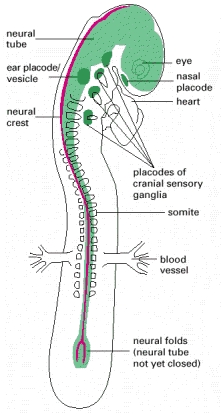
Diagram of a 2 day old embryo illustrating the beginnings of neural development. Where the neural tube (green) is closed and the neural crest (red) is dorsal from ectoderm/above neural tube. [1]
Copyright © 2002, Bruce Alberts, Alexander Johnson, Julian Lewis, Martin Raff, Keith Roberts, and Peter Walter; Copyright © 1983, 1989, 1994, Bruce Alberts, Dennis Bray, Julian Lewis, Martin Raff, Keith Roberts, and James D. Watson . NCBI Bookshelf. A service of the National Library of Medicine, National Institutes of Health.
File history
Click on a date/time to view the file as it appeared at that time.
| Date/Time | Thumbnail | Dimensions | User | Comment | |
|---|---|---|---|---|---|
| current | 16:37, 14 September 2017 | | 221 × 414 (65 KB) | Z5114433 (talk | contribs) | Diagram of 2 day old embryo. The neural tube (green) is closed and the neural crest (red) is dorsal from ectoderm (above neural tube). |
You cannot overwrite this file.
File usage
There are no pages that use this file.