Category:Guinea Pig
From Embryology
This Embryology category shows pages and media related to guinea pig development.
Pages in category 'Guinea Pig'
The following 28 pages are in this category, out of 28 total.
P
- Template:Papanicolaou1933 guinea pig and rat vaginal smear table
- Paper - A contribution to the early development of the heart in mammalia, with special reference to the guinea pig
- Paper - Changes in the vaginal epithelium of the guinea-pig during the oestrous cycle (1922)
- Paper - Growth of the reproductive and endocrine organs of the guinea-pig (1936)
- Paper - Guinea pig development 11 to 21 days
- Paper - Guinea pig development 21 to 35 days
- Paper - Studies on guinea pig oocytes 1
- Paper - The embedding of the embryo guinea-pig in the uterine wall and its nutrition at that stage of development
- Paper - The existence of a typical oestrous cycle in the guinea-pig (1917)
R
- Template:Ref-AdamsHertig1964
- Template:Ref-DeaneslyRowlands1936
- Template:Ref-Emrys-Roberts1910
- Template:Ref-Harman1932
- Template:Ref-Harman1933
- Template:Ref-Huber1918
- Template:Ref-League1928
- Template:Ref-Marion1913
- Template:Ref-Nicol1933
- Template:Ref-Selle1922
- Template:Ref-Silva2016
- Template:Ref-Stockard1917
- Template:Ref-Wilson1928
- Template:Ref-Yoshinaga1921
Media in category 'Guinea Pig'
The following 12 files are in this category, out of 12 total.
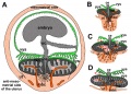 Fetal membrane and placenta cartoon.jpg 600 × 429; 125 KB
 Guineapig icon.jpg 240 × 180; 7 KB
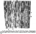 Keibel Mall 332.jpg 851 × 800; 142 KB
 Placenta humans and guinea-pig cartoon.jpg 1,200 × 889; 562 KB
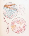
 Placental trophospongium.jpg 567 × 344; 94 KB
 Salvi1898 fig01-15.jpg 2,500 × 1,836; 535 KB
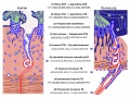
 Skeletal muscle histology 004.jpg 1,280 × 1,024; 242 KB
 Skeletal muscle histology 444.jpg 934 × 701; 125 KB
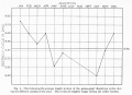 Stockard Papanicolaou1917 figA.jpg 1,382 × 1,000; 143 KB
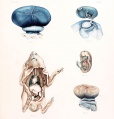 Wislocki1920 plate 1.jpg 1,145 × 1,200; 173 KB
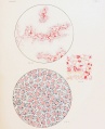
 Wislocki1920 plate 2.jpg 988 × 1,200; 245 KB
 Wislocki1920 plate 3.jpg 975 × 1,200; 219 KB


