Placenta - Histology: Difference between revisions
mNo edit summary |
|||
| (35 intermediate revisions by the same user not shown) | |||
| Line 1: | Line 1: | ||
{{Header}} | |||
==Introduction== | ==Introduction== | ||
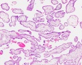
[[File:Placental_villi_6.jpg|thumb|300px|Placental villi with RBC detail.]] | |||
This page introduces the histology of the placenta, the placental cord and fetal membranes. | |||
Various developmental stages of the placental villi are shown including maternal decidua. | |||
{{ | There are images of placental cord and the placental vessels (vein and arteries). | ||
{{Placenta Links}} | |||
{{Histology Links}} | {{Histology Links}} | ||
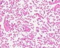
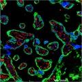
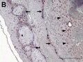
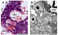
==Placental | ==Hofbauer Cells== | ||
[[File:Placenta Hofbauer cells 01.jpg|thumb|Placental villi Hofbauer cells<ref><pubmed>22606231</pubmed>| [http://www.plosone.org/article/info%3Adoi%2F10.1371%2Fjournal.pone.0035232 PLoS One.]</ref>]] | |||
Hofbauer (1903) identified a sub-population of cells in the placenta that could be identified by specific histological stains, the finding he published 2 years later in 1905.<ref>Hofbauer J. '''About the constant occurrence hitherto unknown cellular form elements in the chorionic villus of the human placenta.''' (''Uber das konstante Vorkommen hisher unbekannter zelliger Formelemente in der Chorionzotte der menschlichen Placenta.'') Wien. klin. Wehnsohr., vol. 16. , 1905. Giundziige einer Biologie der menschlichen Plazenta. Leipzig.</ref> The cells have subsequently been named {{Hofbauer cell}}s, see also the historic 1918 paper by Meyer.<ref>{{Ref-Meyer1918}}</ref> | |||
* human villous macrophages | |||
* highly vacuolated cells | |||
* located the core of placental villi and cord | |||
* macrophages with micropinocytotic activity and phagocytosis ability | |||
* possible paracrine role for early stages of placental vasculogenesis | |||
Search PubMed" [http://www.ncbi.nlm.nih.gov/pubmed/?term=Hofbauer+cells ''Hofbauer cells''] | |||
:'''Historic Links:''' [[Paper - On the nature, occurrence, and identity of the plasma cells of Hofbauer (1918)|1918 plasma cells of Hofbauer]] | [[Book_-_Contributions_to_Embryology_Carnegie_Institution_No.56-14|1921 Early descriptions of Hofbauer cells]] | |||
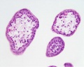
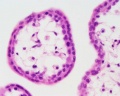
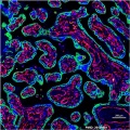
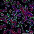
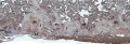
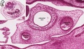
==Villi Histology== | |||
<gallery> | <gallery> | ||
File:Placenta_anchoring_villi.jpg|Placenta anchoring villi and maternal decidua | |||
File:Placental_villi.jpg|First trimester overview | |||
File:Placental_villi_1.jpg|First trimester villi | |||
File:Placental_villi_2.jpg|First trimester detail | |||
File:Placental_villi_5.jpg|Term Overview | |||
File:Placental_villi_3.jpg|Term villi | |||
File:Placental_villi_4.jpg|Term detail | |||
File:Human term placental villi E-cadherin-vimentin.jpg|Term placental villi E-cadherin and vimentin | |||
File:Human term placental villi keratin 7.jpg|Term placental villi keratin 7 | |||
File:Human placental villi keratin 5.jpg|Term placental villi keratin 5 | |||
File:Human placental villi keratin 7.jpg|Term placental villi keratin 7 | |||
File:Human placental villi keratin 8.jpg|Term placental villi keratin 8 | |||
File:Human placental villi keratin 19.jpg|Term placental villi keratin 19 | |||
File:Placenta_histology_006.jpg|Week 15 (GA) | |||
File:Placenta_histology_007.jpg|Week 15 (GA) | |||
File:Placenta_histology_008.jpg | |||
</gallery> | |||
{{Placental villi histology}} | |||
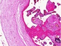
==Cord Histology== | |||
<gallery> | |||
File:Stage 22 image 225.jpg|Week 8 Placental vessels | |||
File:Stage 22 image 203.jpg|Week 8 Placental vessels | |||
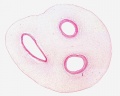
File:Placental cord cross-section.jpg|Placental cord cross-section | File:Placental cord cross-section.jpg|Placental cord cross-section | ||
File:Placental cord cross-section 01.jpg|Placental cord cross-section | |||
File:Placental_vein.jpg|Placental vein | |||
File:Placental_artery_01.jpg|Placental artery | |||
File:Placental_artery.jpg|Placental artery | |||
File:Allantois.jpg|Placental allantois | File:Allantois.jpg|Placental allantois | ||
File:Placental_cord_epithelium_01.jpg|Placental epithelium | File:Placental_cord_epithelium_01.jpg|Placental epithelium | ||
File: | File:Placenta_histology_001.jpg|Placental cord cross-section | ||
File: | File:Placenta_histology_002.jpg|Placental vein | ||
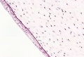
File:Placenta_histology_003.jpg|Whartons jelly | |||
</gallery> | </gallery> | ||
== | {{Placenta Cord Histology}} | ||
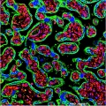
==Blood Histology== | |||
[[File:Placental_villi_6.jpg|800px]] | |||
Placental villi with fetal and maternal red blood cell (RBC) detail. | |||
==Abnormal Histology== | |||
<gallery> | <gallery> | ||
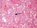
File: | File:Placenta histology 004.jpg|Term placenta | ||
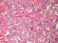
File:Placenta histology 005.jpg|Term placenta chorangiosis | |||
File: | |||
</gallery> | </gallery> | ||
==Other Species Placenta== | |||
== | |||
<gallery> | <gallery> | ||
File: | File:Placental trophospongium.jpg|Guinea Pig related Placenta | ||
</gallery> | </gallery> | ||
==References== | ==References== | ||
Latest revision as of 18:34, 28 December 2019
| Embryology - 2 May 2024 |
|---|
| Google Translate - select your language from the list shown below (this will open a new external page) |
|
العربية | català | 中文 | 中國傳統的 | français | Deutsche | עִברִית | हिंदी | bahasa Indonesia | italiano | 日本語 | 한국어 | မြန်မာ | Pilipino | Polskie | português | ਪੰਜਾਬੀ ਦੇ | Română | русский | Español | Swahili | Svensk | ไทย | Türkçe | اردو | ייִדיש | Tiếng Việt These external translations are automated and may not be accurate. (More? About Translations) |
Introduction
This page introduces the histology of the placenta, the placental cord and fetal membranes.
Various developmental stages of the placental villi are shown including maternal decidua.
There are images of placental cord and the placental vessels (vein and arteries).
| Histology Links: stains | fixatives | artifacts | menstrual histology | placenta histology | heart histology | liver histology | Pancreas | Gall Bladder | Colon | Renal | Respiratory Histology | Bone | Category:Histology | UNSW Histology |
| Historic Histology Textbooks: 1941 Histology] | 1944 Oral Histology |
Hofbauer Cells

Hofbauer (1903) identified a sub-population of cells in the placenta that could be identified by specific histological stains, the finding he published 2 years later in 1905.[2] The cells have subsequently been named Hofbauer cells, see also the historic 1918 paper by Meyer.[3]
- human villous macrophages
- highly vacuolated cells
- located the core of placental villi and cord
- macrophages with micropinocytotic activity and phagocytosis ability
- possible paracrine role for early stages of placental vasculogenesis
Search PubMed" Hofbauer cells
- Historic Links: 1918 plasma cells of Hofbauer | 1921 Early descriptions of Hofbauer cells
Villi Histology

Placenta anchoring villi and maternal decidua
First trimester overview
First trimester villi
First trimester detail
Term Overview
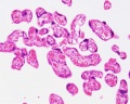
Term villi
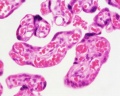
Term detail
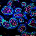
Term placental villi E-cadherin and vimentin
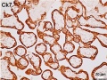
Term placental villi keratin 7
Term placental villi keratin 5
Term placental villi keratin 7
Term placental villi keratin 8
Term placental villi keratin 19
Week 15 (GA)
Week 15 (GA)


- Villi Histology: First trimester (overview | villi | detail) | fetal and maternal RBCs | Term (overview | villi | detail) | Cord Histology | Placenta Histology | Placenta Development
Cord Histology
Week 8 Placental vessels
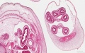
Week 8 Placental vessels

Placental cord cross-section
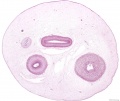
Placental cord cross-section

Placental vein
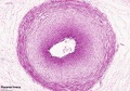
Placental artery

Placental artery

Placental allantois
Placental epithelium
Placental cord cross-section
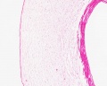
Placental vein
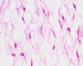
Whartons jelly


- Placental Cord Histology: Cord overview | Vein | Artery | Artery | Allantois | Epithelium | Cord overview 1 unlabeled | overview 2 unlabeled | unlabeled vein and connective tissue | unlabeled connective tissue | Villi histology | Placenta Histology
Blood Histology

Placental villi with fetal and maternal red blood cell (RBC) detail.
Abnormal Histology
Term placenta
Term placenta chorangiosis


Other Species Placenta
Guinea Pig related Placenta

References
- ↑ <pubmed>22606231</pubmed>| PLoS One.
- ↑ Hofbauer J. About the constant occurrence hitherto unknown cellular form elements in the chorionic villus of the human placenta. (Uber das konstante Vorkommen hisher unbekannter zelliger Formelemente in der Chorionzotte der menschlichen Placenta.) Wien. klin. Wehnsohr., vol. 16. , 1905. Giundziige einer Biologie der menschlichen Plazenta. Leipzig.
- ↑ Meyer AW. On the nature, occurrence, and identity of the plasma cells of Hofbauer. (1918) J Morphol. 32(1): 327-350.
Reviews
Articles
Search PubMed
Search Pubmed: Placenta Histology
Glossary Links
- Glossary: A | B | C | D | E | F | G | H | I | J | K | L | M | N | O | P | Q | R | S | T | U | V | W | X | Y | Z | Numbers | Symbols | Term Link
Cite this page: Hill, M.A. (2024, May 2) Embryology Placenta - Histology. Retrieved from https://embryology.med.unsw.edu.au/embryology/index.php/Placenta_-_Histology
- © Dr Mark Hill 2024, UNSW Embryology ISBN: 978 0 7334 2609 4 - UNSW CRICOS Provider Code No. 00098G