File:Artery histology 03.jpg: Difference between revisions
From Embryology
(==Artery Histology== Elastin stain will show: * inner elastic laminae * outer elastic laminae * fine elastic fibres in the tunica media * coarse elastic fibres between the collagen fibres of the tunica adventitia {{Blood Vessel Histology}} {{Blue His) |
No edit summary |
||
| Line 1: | Line 1: | ||
==Artery Histology== | ==Artery Histology== | ||
The tunica intima of elastic arteries will be thicker than in other arteries. | |||
Elastin stain will show: | Elastin stain will show: | ||
Revision as of 08:09, 12 February 2012
Artery Histology
The tunica intima of elastic arteries will be thicker than in other arteries.
Elastin stain will show:
- inner elastic laminae
- outer elastic laminae
- fine elastic fibres in the tunica media
- coarse elastic fibres between the collagen fibres of the tunica adventitia
Links: Histology | Histology Stains | Blue Histology images copyright Lutz Slomianka 1998-2009. The literary and artistic works on the original Blue Histology website may be reproduced, adapted, published and distributed for non-commercial purposes. See also the page Histology Stains.
Cite this page: Hill, M.A. (2024, May 19) Embryology Artery histology 03.jpg. Retrieved from https://embryology.med.unsw.edu.au/embryology/index.php/File:Artery_histology_03.jpg
- © Dr Mark Hill 2024, UNSW Embryology ISBN: 978 0 7334 2609 4 - UNSW CRICOS Provider Code No. 00098G
Aty11he.jpg
File history
Click on a date/time to view the file as it appeared at that time.
| Date/Time | Thumbnail | Dimensions | User | Comment | |
|---|---|---|---|---|---|
| current | 08:05, 12 February 2012 | 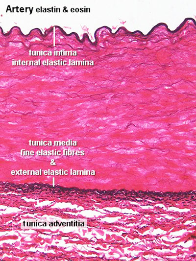 | 400 × 533 (89 KB) | S8600021 (talk | contribs) | ==Artery Histology== Elastin stain will show: * inner elastic laminae * outer elastic laminae * fine elastic fibres in the tunica media * coarse elastic fibres between the collagen fibres of the tunica adventitia {{Blood Vessel Histology}} {{Blue His |
You cannot overwrite this file.
File usage
The following 4 pages use this file: