Category:Blood Vessel
From Embryology
The pages and media listed below relate to the topic of development of blood vessels, both artery and vein.
Note development of lymphatic vessels have a separate category under immune.
Pages in category 'Blood Vessel'
The following 43 pages are in this category, out of 43 total.
C
- Template:Capillary
- Template:CapillaryEM links
- Cardiovascular - Arterial Development
- Cardiovascular - Venous Development
- Cardiovascular System - Blood Vessel Development
- Cardiovascular System - Ductus Arteriosus
- Cardiovascular System - Ductus Venosus
- Cardiovascular System - Foramen Ovale
- Computed Tomography
P
- Paper - Development of the inferior vena cava (1929)
- Paper - On the development of the aortae cardinal and umbilical veins and the other blood vessels of vertebrate embryos from capillaries (1909)
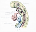
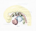
- Paper - On the development of the blood-vessels of the brain in the human embryo (1905)
- Paper - On the origin of the pulmonary arteries in mammals
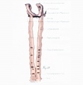
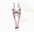
- Paper - The development of the principal arterial stems in the forelimb of the pig (1922)
- Paper - The development of the subcutaneous vascular plexus in the head of the human embryo (1923)
- Paper - The development of the vena cava inferior in man (1925)
- Paper - The origin of the heart and blood vessels in felis domestica (1924)
- Paper The development of the subcutaneous vascular plexus in the head of the human embryo (1923)
R
- Template:Ref-BacsichSmout1938
- Template:Ref-Bremer1902
- Template:Ref-Bremer1909
- Template:Ref-Clark1899b
- Template:Ref-Espinasse1933
- Template:Ref-Gladstone1929
- Template:Ref-Huntington1910a
- Template:Ref-Mall1905
- Template:Ref-McClure1925
- Template:Ref-Miller1903
- Template:Ref-Sabin1908
- Template:Ref-Stockard1915b
- Template:Ref-Wahl1915
- Template:Ref-Watson1924
- Renal System - Abnormalities
- Template:Renal Vascular Anomalies
Media in category 'Blood Vessel'
The following 116 files are in this category, out of 116 total.
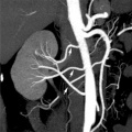
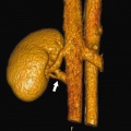
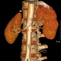 Accessory renal artery.jpg 800 × 798; 103 KB
 Aortic arch branching pattern abnormalities.jpg 656 × 1,404; 133 KB
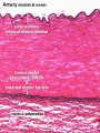
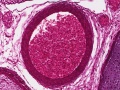
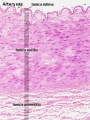 Artery histology 01.jpg 400 × 533; 80 KB
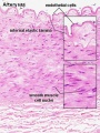 Artery histology 02.jpg 400 × 533; 78 KB
 Artery histology 03.jpg 400 × 533; 89 KB
 Artery histology 04.jpg 800 × 1,000; 93 KB
 Artery histology 05.jpg 400 × 533; 72 KB
 Artery histology 06.jpg 400 × 533; 91 KB
 Artery histology 11.jpg 1,280 × 1,024; 344 KB
 Artery histology 12.jpg 1,280 × 1,024; 206 KB
 Artery histology 13.jpg 1,280 × 1,024; 474 KB
 Artery histology 14.jpg 1,280 × 1,024; 466 KB
 Artery histology 15.jpg 1,280 × 1,024; 343 KB
 Artery histology 16.jpg 1,280 × 1,024; 409 KB
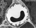
 Blood capillary EM 01.jpg 1,107 × 714; 260 KB
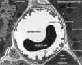
 Blood capillary EM 02.jpg 600 × 600; 99 KB
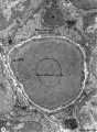
 Blood capillary EM 03.jpg 1,560 × 1,230; 441 KB
 Blood capillary EM 04.jpg 1,560 × 1,230; 462 KB
 Blood capillary EM 05.jpg 1,015 × 800; 205 KB
 Blood capillary EM 06.jpg 1,015 × 800; 216 KB
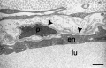
 Blood-thymus barrier EM01.jpg 1,280 × 1,747; 375 KB
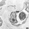
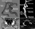
 Cerebral blood supply development 01.jpg 1,200 × 460; 67 KB
 Congdon1922-1-16.jpg 980 × 1,000; 157 KB
 Congdon1922-17.jpg 1,000 × 411; 55 KB
 Congdon1922-18-25.jpg 1,200 × 795; 179 KB
 Congdon1922-18.jpg 494 × 506; 29 KB
 Congdon1922-19.jpg 653 × 471; 31 KB
 Congdon1922-20.jpg 794 × 446; 43 KB
 Congdon1922-21.jpg 578 × 407; 27 KB
 Congdon1922-22.jpg 511 × 489; 27 KB
 Congdon1922-23.jpg 519 × 412; 26 KB
 Congdon1922-24.jpg 790 × 482; 40 KB
 Congdon1922-25.jpg 509 × 358; 25 KB
 Congdon1922-26.jpg 746 × 726; 54 KB
 Congdon1922-27-28.jpg 997 × 612; 68 KB
 Congdon1922-29.jpg 976 × 1,000; 81 KB
 Congdon1922-30.jpg 1,133 × 1,000; 176 KB
 Congdon1922-31.jpg 1,063 × 1,000; 93 KB
 Congdon1922-32.jpg 1,133 × 1,000; 132 KB
 Congdon1922-33.jpg 920 × 1,000; 107 KB
 Congdon1922-34.jpg 920 × 1,000; 122 KB
 Congdon1922-35.jpg 920 × 1,000; 97 KB
 Congdon1922-36.jpg 920 × 1,000; 113 KB
 Congdon1922-37.jpg 1,200 × 838; 163 KB
 Congdon1922-38.jpg 1,187 × 1,000; 165 KB
 Congdon1922-39.jpg 1,200 × 756; 138 KB
 Congdon1922-40.jpg 1,013 × 1,000; 110 KB
 Congdon1922-plate01.jpg 877 × 1,200; 145 KB
 Congdon1922-plate02.jpg 877 × 1,200; 191 KB
 Congdon1922-plate03.jpg 1,200 × 885; 182 KB
 Finley1923 fig01.jpg 494 × 968; 53 KB
 Finley1923 fig02.jpg 700 × 800; 77 KB
 Finley1923 fig03.jpg 600 × 512; 56 KB
 Finley1923 fig04.jpg 700 × 627; 61 KB
 Finley1923 fig05.jpg 674 × 800; 146 KB
 Finley1923 fig06.jpg 729 × 800; 95 KB
 Finley1923 fig07.jpg 314 × 800; 40 KB
 Finley1923 fig08.jpg 296 × 800; 32 KB
 Finley1923 fig09.jpg 456 × 800; 49 KB
 Finley1923 fig10.jpg 221 × 802; 17 KB
 Finley1923 fig11.jpg 432 × 800; 38 KB
 Finley1923 fig12.jpg 593 × 800; 48 KB
 Finley1923 fig13.jpg 594 × 800; 51 KB
 Finley1923 Plate 1.jpg 776 × 1,000; 151 KB
 Finley1923 Plate 2.jpg 864 × 1,200; 153 KB
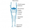
 Gray0448.jpg 356 × 600; 70 KB
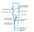
 Gray0460.jpg 818 × 551; 106 KB
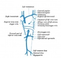
 Gray0461.jpg 859 × 841; 82 KB
 Gray0462.jpg 822 × 800; 157 KB
 Gray0463.jpg 1,099 × 755; 134 KB
 Gray0464.jpg 993 × 961; 258 KB
 Gray0465.jpg 954 × 938; 207 KB
 Gray0473.jpg 499 × 418; 0 bytes
 Gray0477.jpg 597 × 560; 22 KB
 Gray0478.jpg 597 × 560; 21 KB
 Gray0479.jpg 597 × 560; 26 KB
 Gray0480.jpg 597 × 560; 31 KB
 McClure1925 fig01.jpg 1,000 × 1,585; 116 KB
 McClure1925 fig02.jpg 1,000 × 1,364; 159 KB
 McClure1925 fig03.jpg 1,000 × 1,563; 150 KB
 McClure1925 fig04.jpg 1,000 × 1,480; 258 KB
 McClure1925 fig05.jpg 1,000 × 1,548; 151 KB
 McClure1925 fig06.jpg 922 × 1,227; 196 KB
 McClure1925 fig07.jpg 1,000 × 1,509; 185 KB
 McClure1925 fig08.jpg 900 × 1,245; 193 KB
 McClure1925 fig09.jpg 1,000 × 1,474; 193 KB
 McClure1925 fig10.jpg 1,000 × 1,437; 311 KB
 McClure1925 fig11.jpg 900 × 1,047; 185 KB
 McClure1925 fig12.jpg 1,000 × 1,564; 184 KB
 McClure1925 fig13.jpg 1,000 × 1,368; 331 KB
 McClure1925 fig14.jpg 1,000 × 1,586; 211 KB
 McClure1925 fig15.jpg 1,000 × 1,301; 309 KB
 McClure1925 fig16.jpg 1,000 × 919; 212 KB
 McClure1925 fig17.jpg 1,000 × 1,470; 197 KB
 McClure1925 fig18.jpg 1,000 × 1,401; 158 KB
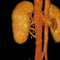
 Multiple renal arteries 01.jpg 496 × 496; 40 KB
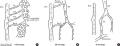
 Notch and yolk sac blood vessels model.jpg 600 × 775; 97 KB
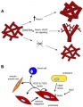
 NOTCH-endothelial-cartoon.jpg 1,280 × 888; 92 KB
 Stage 22 image 223.jpg 1,128 × 846; 366 KB
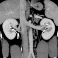
 Supernumerary renal vein 01.jpg 800 × 798; 72 KB
 Supernumerary renal vein 02.jpg 800 × 795; 89 KB
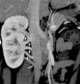
 Supernumerary renal vein 03.jpg 800 × 794; 80 KB
 Supernumerary renal vein 04.jpg 800 × 850; 76 KB
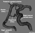
 Trigeminal artery 01.jpg 947 × 800; 102 KB
 Trigeminal artery 02.jpg 520 × 490; 35 KB
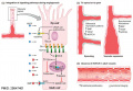
 Vasculature development 01 cartoon.jpg 599 × 900; 106 KB
 Vasculature development 02 cartoon.jpg 601 × 1,002; 93 KB
 Vein valve animation.gif 300 × 200; 54 KB
 Venule microvessel EM.jpg 600 × 626; 91 KB
 Woollard-plate01.jpg 744 × 1,000; 153 KB
 Woollard-plate02.jpg 788 × 1,000; 196 KB
 Woollard001.jpg 739 × 858; 142 KB
 Woollard002.jpg 699 × 873; 125 KB
 Woollard003.jpg 1,107 × 848; 159 KB
 Woollard004.jpg 1,037 × 717; 148 KB
 Woollard005.jpg 1,034 × 655; 144 KB


