Lecture - Week 1 and 2 Development
2010 Lecture | Zygote | Blastocyst | Implantation
Objectives
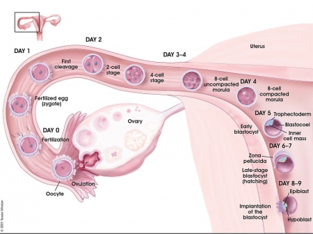
- Understand the events during week 1 of development (Zygote, Blastomeres, Morula, Blastocyst)
- Understand the events during week 2 of development (Trophoblast, Syncytiotrophoblast, Cytotrophoblast, Embryoblast, Implantation)
- Brief understanding of early placentation
- Brief understanding of maternal changes
Zygote Formation
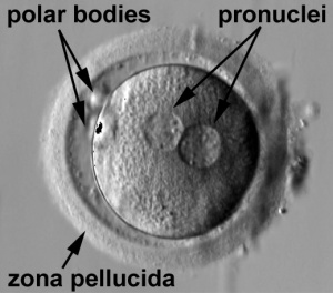
- zygote is the first diploid cell formed following fertilisation.
- male and female pronuclei, 2 nuclei approach each other and nuclear membranes break down.
- DNA replicates, first mitotic division
- sperm contributes centriole which organizes mitotic spindle
|
Movie - Pronuclear Fusion | Movie - Parental Genomes
- Conceptus - the term refers to all material derived from this fertilised zygote, includes both the embryo and the non-embryonic tissues (placenta, fetal membranes).
- Links: Carnegie stage 1
Cleavage of Zygote
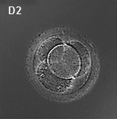
- cleavage of zygote forms 2 blastomeres and is also cleavage with no cytoplasm synthesis.
- special "embryonic" cell cycle S phases and M phases alternate without any intervening G1 or G2 phases (MSMSMSMS, adult MG1SG2) therefore individual cell volume decreases.
- cell division is initially synchronous, then asynchronously
- slow- centre cells, larger fast- peripheral cells
- zona pellucid still intact (division occurs within the ZP)
<wikiflv width="414" height="278" autoplay="true">embryo mitosis.flv|Embryo_mitosis_icon.jpg</wikiflv>
Morula
|
|
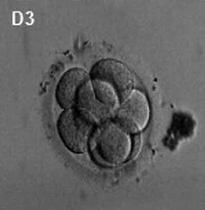
| Human Embryo (day 2) | Human Embryo (day 3) |
- about day 4 is a solid ball of 16-20 cells with peripheral cells flattened against zona pellucida
- compaction occurs forming a cavity and leading to the next blastocyst stage
Blastocyst
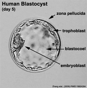
- about day 5 have 2 identifiable cell types and a fluid-filled cavity (blastoceol)
- outer cell layer - trophoblast, peripheral flattened cells, forms the placenta and placental membranes
- inner cell mass - embryoblast, mass of rounder cells located on one wall of the blastocoel, forms entire embryo
Blastula Cell Communication
Two forms of cellular junctions Junctions
- gap junctions, allow electrically couple cells of epithelium surrounding a fluid-filled cavity
- tight junctions, close to outer surface create a seal, isolates interior of embryo from external medium
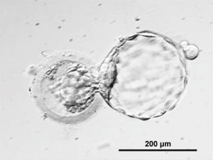
Blastocyst Hatching
Blastocyst Hatching - zona pellucida lost, ZP has sperm entry site, and entire ZP broken down by uterine secretions and possibly blastula secretions. Uterine Glands - secretions required for blastocyst motility and nutrition
Week 2 - Implantation
The second week of human development is concerned with the process of implantation and the differentiation of the blastocyst into early embryonic and placental forming structures.
Normal Implantation Sites - in uterine wall superior, posterior, lateral |
File:Week2_001_icon.jpg</wikiflv> |
Endometrial Receptivity
- In humans, receptivity occurs 6 days after the post-ovulatory progesterone surge and lasts about 2 to 4 days.
- Similar "receptivity window" in other species (rat day 5 and mouse day 4.5).
- Many studies have looked into identifying markers for this receptivity period both to optimise and to block this process.
Abnormal Implantation
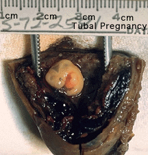
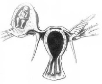
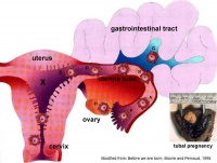
Abnormal implantation sites or Ectopic Pregnancy occurs if implantation is in uterine tube or outside the uterus.
- sites - external surface of uterus, ovary, bowel, gastrointestinal tract, mesentry, peritoneal wall
- If not spontaneous then, embryo has to be removed surgically
Tubal pregnancy - 94% of ectopic pregnancies
- if uterine epithelium is damaged (scarring, pelvic inflammatory disease)
- if zona pellucida is lost too early, allows premature tubal implantation
- embryo may develop through early stages, can erode through the uterine horn and reattach within the peritoneal cavity
|
|
Co-ordinator Note
Dr Mark Hill |
ANAT2341 Embryology S2 2011
|
Course Content 2011
2011 Timetable: | Embryology Introduction | Fertilization | Cell Division/Fertilization | Week 1 and 2 Development | Week 3 Development | Week 1 to 3 | Mesoderm Development | Ectoderm, Early Neural, Neural Crest | Trilaminar Embryo to Early Embryo | Early Vascular Development | Placenta | Vascular and Placenta | Endoderm, Early Gastrointestinal | Respiratory Development | Endoderm and Respiratory | Head Development | Neural Crest Development | Head and Neural Crest | Musculoskeletal Development | Limb Development | Musculoskeletal | Renal Development | Genital | Kidney and Genital | Sensory | Stem Cells | Stem Cells | Endocrine Development | Endocrine | Heart | Integumentary Development | Heart and Integumentary | Fetal | Birth and Revision | Fetal
Glossary Links
- Glossary: A | B | C | D | E | F | G | H | I | J | K | L | M | N | O | P | Q | R | S | T | U | V | W | X | Y | Z | Numbers | Symbols | Term Link
Cite this page: Hill, M.A. (2024, May 23) Embryology Lecture - Week 1 and 2 Development. Retrieved from https://embryology.med.unsw.edu.au/embryology/index.php/Lecture_-_Week_1_and_2_Development
- © Dr Mark Hill 2024, UNSW Embryology ISBN: 978 0 7334 2609 4 - UNSW CRICOS Provider Code No. 00098G
