Category:Histology
From Embryology
Introduction
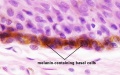
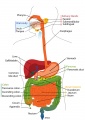
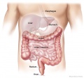
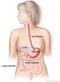
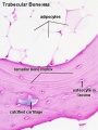
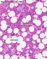
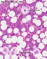
Files and media below relate to histology. Most links are to histological images that relate to tissue structure and development. Many images are sourced from the original UNSW Anatomy Histology slide set and UWA Blue Histology online images.
Subcategories
This category has the following 15 subcategories, out of 15 total.
Pages in category 'Histology'
The following 200 pages are in this category, out of 334 total.
(previous page) (next page)A
- Template:Adrenal Histology
- AE Practical - Neural Histology
- Template:Al. coch.
- Alagille Syndrome
- ANAT2241 Blood
- ANAT2241 Bone, Bone Formation and Joints
- ANAT2241 Cardiovascular System
- ANAT2241 Connective Tissue Components
- ANAT2241 Connective Tissue Types
- ANAT2241 Covering and Lining Epithelia
- ANAT2241 Endocrine System
- ANAT2241 Female Reproductive System
- Template:ANAT2241 footer
- ANAT2241 Gastro-Intestinal System
- ANAT2241 Glandular Epithelia
- ANAT2241 Histology - Basic and Systematic
- Talk:ANAT2241 Histology - Basic and Systematic
- ANAT2241 Integumentary System
- ANAT2241 Liver, Gallbladder, and Pancreas
- ANAT2241 Lymphatic Tissue and Immune System
- ANAT2241 Male Reproductive System
- ANAT2241 Muscle Tissue
- ANAT2241 Nervous Tissue
- ANAT2241 Respiratory System
- ANAT2241 Special Senses The Eye
- ANAT2241 The Virtual Microscope
- ANAT2241 Urinary System
- ANAT2241 Virtual Slides - Example Spot Images
- ANAT2511 - Fundamentals of Anatomy
- ANAT2511 Basic Tissues
- ANAT2511 Bones and Joints
- ANAT2511 Circulatory System
- Template:ANAT2511 footer
- ANAT2511 Gastrointestinal Tract
- ANAT2511 Integumentary System
- ANAT2511 Introduction to Histology
- ANAT2511 Muscle Tissue
- ANAT2511 Nervous Tissue
- ANAT2511 Respiratory System
- ANAT2511 Urinary System
- Template:Artifacts
B
- BGD Lecture - Endocrine Histology
- BGD Lecture - Gastrointestinal System Development
- Talk:BGD Lecture - Gastrointestinal System Development
- BGDA Practical - Female Reproductive Tract Histology
- BGDB Gastrointestinal - Abnormalities
- BGDB Practical - Gastrointestinal System Development
- Talk:BGDB Practical - Gastrointestinal System Development
- BGDB Practical - Upper Gastrointestinal Tract Histology
- Blood Cell Histology Movie
- Template:Blood Histology
- Template:Blood Vessel Histology
- Template:Blood vessel histology
- Template:Blue Histology
- Bone Development
- Bone Histology
- Template:Bone Histology
- Template:Bone histology
- Template:Bone Marrow Histology
- Book - A laboratory guide in histology (1917)
- Book - A Laboratory Text-Book of Embryology 8 (1903)
- Book - A text-book of histology arranged upon an embryological basis (1913)
- Book - A text-book of histology arranged upon an embryological basis (1913) 1
- Book - A text-book of histology arranged upon an embryological basis (1913) 1-1
- Book - A text-book of histology arranged upon an embryological basis (1913) 1-2
- Book - A text-book of histology arranged upon an embryological basis (1913) 1-3
- Book - A text-book of histology arranged upon an embryological basis (1913) 2
- Book - A text-book of histology arranged upon an embryological basis (1913) 2-1
- Book - A text-book of histology arranged upon an embryological basis (1913) 2-2
- Book - A textbook of histology, including microscopic technic (1910) General Histology 1
- Book - A textbook of histology, including microscopic technic (1910) General Histology 2
- Book - A textbook of histology, including microscopic technic (1910) microscopic technic
- Book - A textbook of histology, including microscopic technic (1910) Special Histology 1
- Book - A textbook of histology, including microscopic technic (1910) Special Histology 10
- Book - A textbook of histology, including microscopic technic (1910) Special Histology 2
- Book - A textbook of histology, including microscopic technic (1910) Special Histology 3
- Book - A textbook of histology, including microscopic technic (1910) Special Histology 4
- Book - A textbook of histology, including microscopic technic (1910) Special Histology 5
- Book - A textbook of histology, including microscopic technic (1910) Special Histology 6
- Book - A textbook of histology, including microscopic technic (1910) Special Histology 7
- Book - A textbook of histology, including microscopic technic (1910) Special Histology 8
- Book - A textbook of histology, including microscopic technic (1910) Special Histology 9
- Book - Contributions to Embryology Carnegie Institution No.50
- Book - Histology and Embryology 1941
- Book - Liver development
- Book - Stoehr's Histology (1906)
- Book - Stoehr's Histology 1
- Talk:Book - Stoehr's Histology 1
- Book - Stoehr's Histology 1-1
- Book - Stoehr's Histology 1-2
- Book - Stoehr's Histology 1-3
- Book - Stoehr's Histology 2
- Book - Stoehr's Histology Figures
- Book - The microscopic anatomy of the human body, in health and disease
- Template:Bouin
- Buccopharyngeal membrane
- Template:BöhmDavidoffHuber1910 footer
- Template:BöhmDavidoffHuber1910 TOC
C
- Template:CapillaryEM links
- Template:Cardiac muscle EM
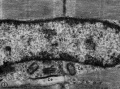
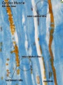
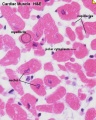
- Cardiac Muscle Histology
- Cardiovascular - Arterial Development
- Cardiovascular System - Blood Development
- Cardiovascular System - Heart Histology
- Cardiovascular System - Spleen Development
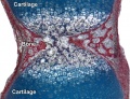
- Cartilage Histology
- Template:Cartilage Histology
- Template:Cartilage histology
- Cloaca Development
- Colon Histology 2009
- Template:Common Stains collapsetable
- Template:Common Stains table
E
F
G
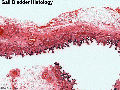
- Template:Gall Bladder Histology Images
- Gastrointestinal Tract - Abnormalities
- Gastrointestinal Tract - Carnegie Stage 13
- Gastrointestinal Tract - Carnegie Stage 22
- Gastrointestinal Tract - Colon Histology
- Gastrointestinal Tract - Gall Bladder Development
- Gastrointestinal Tract - Gall Bladder Histology
- Gastrointestinal Tract - Gallbladder Development
- Gastrointestinal Tract - Gallbladder Histology
- Gastrointestinal Tract - Histology
- Gastrointestinal Tract - Intestine Development
- Gastrointestinal Tract - Liver Development
- Gastrointestinal Tract - Liver Histology
- Gastrointestinal Tract - Mesentery Development
- Gastrointestinal Tract - Mouth Development
- Gastrointestinal Tract - Oesophagus Development
- Gastrointestinal Tract - Pancreas Development
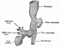
- Gastrointestinal Tract - Pancreas Histology
- Gastrointestinal Tract - Postnatal
- Gastrointestinal Tract - Stomach Development
- Gastrointestinal Tract Development
- Template:Gastrointestinal Tract Links
- Template:GIT histology links
H
- Template:Hair histology links
- Template:HE
- Template:Heart histology
- Template:HillH52
- Histology
- Template:Histology
- Histology and Embryology 1941 - Bibliography
- Histology and Embryology 1941 - Embryology
- Histology and Embryology 1941 - Embryology 1
- Histology and Embryology 1941 - Embryology 2
- Histology and Embryology 1941 - Histology
- Histology and Embryology 1941 - Histology 1
- Histology and Embryology 1941 - Histology 2
- Histology and Embryology 1941 - Histology 3
- Histology Artifacts
- Histology Fixatives
- Template:Histology Links
- Histology Stains
- Template:Histology Stains
- HM Practical - Blood Vessel Histology
- HM Practical - Cardiac Histology
- Template:Human follicles lm and em links
- Template:Human ovary - corpus luteum links
- Human System Development
I
L
M
P
- Template:Pancreas Histology Images
- Paper - A case of atresia ani in a human embryo of 26 mm
- Paper - A case of atresia of the esophagus combined with traoheoesophageal fistula in a 9 mm human embryo, and its embryological explanation
- Paper - A case of congenital malformations of the intestinal canal (1923)
- Paper - A Contribution to the Embryology of the Liver and Vascular System in Man
- Paper - A contribution to the morphology and development of the mammalian liver
- Paper - A contribution to the morphology and development of the mammalian liver (1908)
- Paper - A histological investigation of the development and structure of the human lung
- Paper - A model demonstrating the changes in position and peritoneal relations of abdominal viscera during development (1912)
Media in category 'Histology'
The following 200 files are in this category, out of 718 total.
(previous page) (next page)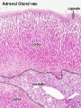 Adrenal histology 001.jpg 450 × 600; 151 KB
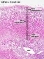 Adrenal histology 002.jpg 450 × 600; 150 KB
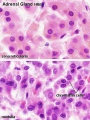 Adrenal histology 003.jpg 450 × 600; 71 KB
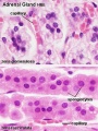 Adrenal histology 004.jpg 450 × 600; 71 KB
 Adrenal histology 005.jpg 1,280 × 1,024; 298 KB
 Adrenal histology 006.jpg 1,280 × 1,024; 313 KB
 Adrenal histology 007.jpg 1,280 × 1,024; 298 KB
 Adrenal histology 008.jpg 1,280 × 1,024; 272 KB
 Adrenal histology 009.jpg 1,280 × 1,024; 265 KB
 Adrenal histology 010.jpg 1,280 × 1,024; 292 KB
 Adrenal histology 011.jpg 1,280 × 1,024; 572 KB
 Adult epidermis histology 01.jpg 600 × 750; 83 KB
 Adult epidermis histology 02.jpg 600 × 750; 123 KB
 Adult epidermis histology 03.jpg 600 × 375; 46 KB
 Adult gastrointestinal tract cartoon.jpg 707 × 1,000; 105 KB
 Adult gastrointestinal tract cartoon01.jpg 745 × 698; 55 KB
 Adult gastrointestinal tract cartoon02.jpg 541 × 738; 42 KB
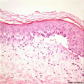
 Adult skin histology 01.jpg 600 × 750; 73 KB
 Adult skin histology 02.jpg 600 × 750; 88 KB
 Adult skin histology 03.jpg 600 × 750; 108 KB
 Adult skin histology 04.jpg 480 × 600; 128 KB
 Allantois.jpg 600 × 531; 51 KB
 ANAT2241 Epithelia Lecture2018 4 slides.pdf ; 16.08 MB
- ANAT2241 Epithelia Lecture2018.pdf ; 17.51 MB
 Apocrine secretion animation.gif 60 × 80; 4 KB
 ARecVG02.jpg 300 × 400; 113 KB
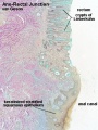
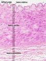 Artery histology 01.jpg 400 × 533; 80 KB
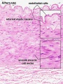 Artery histology 02.jpg 400 × 533; 78 KB
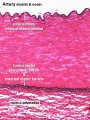 Artery histology 03.jpg 400 × 533; 89 KB
 Artery histology 04.jpg 800 × 1,000; 93 KB
 Artery histology 05.jpg 400 × 533; 72 KB
 Artery histology 06.jpg 400 × 533; 91 KB
 Artery histology 11.jpg 1,280 × 1,024; 344 KB
 Artery histology 12.jpg 1,280 × 1,024; 206 KB
 Artery histology 13.jpg 1,280 × 1,024; 474 KB
 Artery histology 14.jpg 1,280 × 1,024; 466 KB
 Artery histology 15.jpg 1,280 × 1,024; 343 KB
 Artery histology 16.jpg 1,280 × 1,024; 409 KB
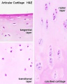 Articular cartilage 01.jpg 500 × 626; 42 KB
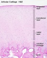 Articular cartilage.jpg 500 × 626; 77 KB
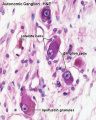 Autonomic ganglion histology 01.jpg 641 × 800; 56 KB
 Azoospermia.jpg 768 × 554; 77 KB
 Bacteria - gram-stained vaginal smear 01.jpg 1,000 × 749; 185 KB
 Bacteria - gram-stained vaginal smear 02.jpg 1,000 × 749; 219 KB
 Bacteria - gram-stained vaginal smear 03.jpg 1,000 × 749; 186 KB
 Bacteria - gram-stained vaginal smear 04.jpg 1,000 × 749; 174 KB
 Bacteria - gram-stained vaginal smear 05.jpg 1,000 × 749; 201 KB
 Bacteria - gram-stained vaginal smear 06.jpg 1,000 × 749; 194 KB
 Bacteria - gram-stained vaginal smear 07.jpg 1,000 × 749; 216 KB
 Bacteria - gram-stained vaginal smear 08.jpg 1,000 × 749; 180 KB
 Bacteria - gram-stained vaginal smear 09.jpg 1,000 × 749; 176 KB
 Bacteria - gram-stained vaginal smear 10.jpg 1,000 × 749; 237 KB
 Bacteria - gram-stained vaginal smear 11.jpg 1,000 × 749; 224 KB
 Bacteria - gram-stained vaginal smear 12.jpg 1,000 × 749; 177 KB
 Bailey274.jpg 558 × 442; 32 KB
 Basophil 01.jpg 480 × 600; 37 KB
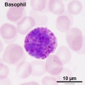 Basophil 02.jpg 600 × 600; 44 KB
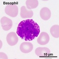 Basophil 03.jpg 600 × 600; 51 KB
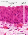
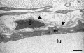
 Bladder histology 001.jpg 1,280 × 1,024; 522 KB
 Bladder histology 002.jpg 1,280 × 1,024; 295 KB
 Bladder histology 003.jpg 1,280 × 1,024; 229 KB
 Bladder histology 004.jpg 1,280 × 1,024; 212 KB
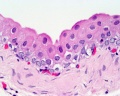
 Bladder histology 01.jpg 480 × 600; 29 KB
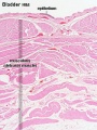 Bladder histology.jpg 300 × 400; 56 KB
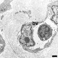
 Blood capillary EM 01.jpg 1,107 × 714; 260 KB
 Blood capillary EM 02.jpg 600 × 600; 99 KB
 Blood capillary EM 03.jpg 1,560 × 1,230; 441 KB
 Blood capillary EM 04.jpg 1,560 × 1,230; 462 KB
 Blood capillary EM 05.jpg 1,015 × 800; 205 KB
 Blood capillary EM 06.jpg 1,015 × 800; 216 KB
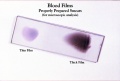 Blood Smear Slide 01.jpg 700 × 471; 58 KB
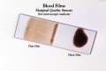 Blood Smear Slide 02.jpg 700 × 471; 47 KB
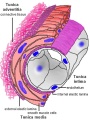 Blood vessel wall cartoon.jpg 450 × 600; 71 KB
 Bone histology 001.jpg 1,280 × 1,024; 276 KB
 Bone histology 002.jpg 1,280 × 1,024; 309 KB
 Bone histology 003.jpg 1,280 × 1,024; 663 KB
 Bone histology 004.jpg 1,280 × 1,024; 605 KB
 Bone histology 005.jpg 1,280 × 1,024; 529 KB
 Bone histology 006.jpg 1,280 × 1,024; 360 KB
 Bone histology 007.jpg 1,280 × 1,024; 299 KB
 Bone histology 008.jpg 1,280 × 1,024; 550 KB
 Bone histology 009.jpg 1,280 × 1,024; 444 KB
 Bone histology 010.jpg 1,280 × 1,024; 256 KB
 Bone histology 011.jpg 1,280 × 1,024; 348 KB
 Bone histology 012.jpg 1,280 × 1,024; 165 KB
 Bone histology 013.jpg 1,280 × 1,024; 210 KB
 Bone histology 014.jpg 1,280 × 1,024; 541 KB
 Bone histology 015.jpg 1,280 × 1,024; 519 KB
 Bone histology 016.jpg 1,280 × 1,024; 379 KB
 Bone histology 017.jpg 1,280 × 1,024; 442 KB
 Bone histology 018.jpg 1,280 × 1,024; 336 KB
 Bone histology 019.jpg 1,280 × 1,024; 275 KB
 Bone histology 020.jpg 1,280 × 1,024; 272 KB
 Bone histology 021.jpg 1,280 × 1,024; 254 KB
 Bone histology 022.jpg 2,500 × 2,000; 328 KB
 Bone histology 066.jpg 2,500 × 2,000; 361 KB
 Bone histology 101.jpg 400 × 533; 59 KB
 Bone histology 111.jpg 400 × 533; 70 KB
 Bone histology 112.jpg 400 × 533; 46 KB
 Bone histology 201.jpg 400 × 533; 62 KB
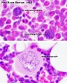
 Bone marrow histology 01.jpg 480 × 600; 114 KB
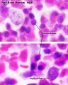
 Bone marrow histology 02.jpg 480 × 600; 109 KB
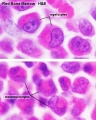
 Bone marrow histology 03.jpg 480 × 600; 81 KB
 Bone marrow histology 04.jpg 480 × 600; 61 KB
 Bone marrow histology 05.jpg 480 × 600; 62 KB
 Bone-bon02he.jpg 1,280 × 1,024; 348 KB
 Bone-femur.jpg 798 × 1,000; 150 KB
 Bone-structure.jpg 450 × 600; 27 KB
 Borrelia-burgdorferi.jpg 285 × 214; 14 KB
 Boyd collection icon.jpg 400 × 554; 56 KB
 Brain histology 01.jpg 480 × 600; 125 KB
 Brain histology 02.jpg 480 × 600; 51 KB
 Brown adipose histology.jpg 400 × 500; 64 KB
 Camillo Golgi.jpg 318 × 450; 39 KB
 Cardiac muscle EM01.jpg 1,072 × 735; 231 KB
 Cardiac muscle EM02.jpg 1,072 × 735; 224 KB
 Cardiac muscle EM03.jpg 849 × 615; 135 KB
 Cardiac muscle EM04.jpg 1,000 × 680; 191 KB
 Cardiac Muscle EM05.jpg 992 × 733; 158 KB
 Cardiac muscle histology.jpg 300 × 400; 42 KB
 Cartilage em01.jpg 800 × 551; 176 KB
 Cartilage histology 001.jpg 1,280 × 1,024; 362 KB
 Cartilage histology 002.jpg 1,280 × 1,024; 174 KB
 Cartilage histology 003.jpg 1,280 × 1,024; 161 KB
 Cartilage histology 004.jpg 1,280 × 1,024; 216 KB
 Cartilage histology 005.jpg 1,280 × 1,024; 344 KB
 Cartilage nest.gif 150 × 200; 154 KB
 Col00he.jpg 1,280 × 1,024; 117 KB
 Colon histology 001.jpg 400 × 533; 64 KB
 Colon histology 002.jpg 300 × 400; 73 KB
 Colon histology 003.jpg 1,280 × 1,024; 117 KB
 Colon histology 004.jpg 1,280 × 1,024; 292 KB
 Colon histology 005.jpg 1,280 × 1,024; 403 KB
 Colon histology 006.jpg 400 × 533; 70 KB
 Colon histology 007.jpg 1,280 × 1,024; 152 KB
 Colon histology 008.jpg 1,278 × 959; 237 KB
 Colon histology 009.jpg 1,280 × 1,024; 159 KB
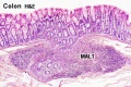 Colon MALT.jpg 500 × 333; 67 KB
 Complete hydatidiform mole 04.jpg 748 × 560; 124 KB
 Complete hydatidiform mole 06.jpg 1,280 × 960; 553 KB
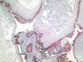
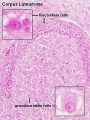 Corpus luteum lutein cells.jpg 450 × 600; 104 KB
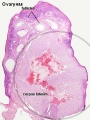 Corpus luteum.jpg 450 × 600; 94 KB
 Danchakoff1916 plate01+overlay.jpg 1,113 × 592; 60 KB
 Danchakoff1916 plate01.jpg 1,113 × 592; 49 KB
 Danchakoff1916 plate01overlay.jpg 1,113 × 592; 81 KB
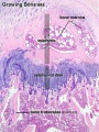
 Developing vertebra.jpg 558 × 428; 93 KB
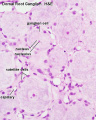 Dorsal root ganglion histology 01.jpg 640 × 800; 45 KB
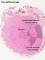 Ductus deferens 01.jpg 400 × 533; 76 KB
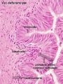 Ductus deferens 02.jpg 400 × 533; 80 KB
 Duodenum cartoon.jpg 500 × 704; 42 KB
 Duodenum histology 01.jpg 480 × 600; 83 KB
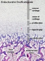
 Endochondral bone.jpg 600 × 451; 89 KB
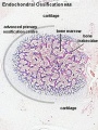 Endochondral ossification 1.jpg 400 × 534; 91 KB
 Endochondral ossification 2.jpg 400 × 533; 99 KB
 Endochondral ossification.jpg 400 × 533; 91 KB
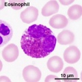
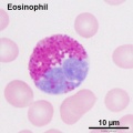 Eosinophil 01.jpg 600 × 600; 49 KB
 Eosinophil 02.jpg 600 × 600; 58 KB
 Epidermolysis bullosa simplex 01.jpg 498 × 498; 62 KB
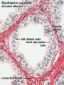
 Epididymis histology 01.jpg 600 × 375; 20 KB
 Epididymis histology 02.jpg 400 × 534; 71 KB
 Epididymis histology 03.jpg 400 × 533; 68 KB
 Epiglottis cartilage 01.jpg 500 × 626; 95 KB
 Epiglottis cartilage 02.jpg 500 × 626; 101 KB
 Epithelium histology cartoon 01.jpg 600 × 376; 23 KB
 Fetal cartilage 01.jpg 639 × 400; 71 KB
 Fetal corpus cavernosum and corpus spongiosum 01.jpg 1,795 × 2,082; 919 KB
 Fetal integumentary histology 01.jpg 800 × 219; 74 KB
 Fetal integumentary histology 02.jpg 600 × 664; 145 KB
 Fetal lung histology 01.jpg 1,280 × 1,024; 339 KB
 Fetal lung histology 02.jpg 450 × 600; 74 KB
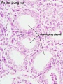 Fetal lung histology.jpg 450 × 600; 83 KB
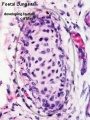
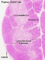 Fetal thymus.jpg 450 × 600; 122 KB
 Fetalblood.jpg 400 × 307; 24 KB
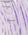 Fibrous cartilage 01.jpg 500 × 626; 76 KB
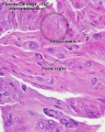 Fibrous cartilage 02.jpg 500 × 626; 96 KB
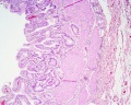
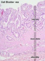 Gall bladder histology 001.jpg 375 × 500; 78 KB
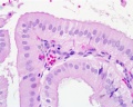
 Gall bladder histology 002.jpg 375 × 500; 45 KB
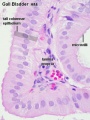
 Gall bladder histology 003.jpg 1,280 × 1,024; 577 KB
 Gall bladder histology 004.jpg 1,280 × 1,024; 254 KB
 Gall bladder histology 005.gif 600 × 450; 683 KB
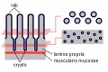 Gastrointestinal villi and crypts cartoon.jpg 500 × 333; 28 KB
 Gray0964.jpg 536 × 800; 139 KB
 Gray0976.jpg 600 × 599; 146 KB
 Hair histology.jpg 600 × 451; 131 KB
 Hassall1849 plate41 fig10.jpg 364 × 1,000; 73 KB
 Hassall1849 plate41.jpg 1,280 × 2,075; 857 KB
 Heart histology 001.jpg 400 × 500; 83 KB
 Heart histology 002.jpg 400 × 500; 81 KB
 Heart histology 003.jpg 400 × 500; 136 KB
 Heart histology 004.jpg 400 × 500; 97 KB
 Heart histology 101.jpg 1,280 × 1,024; 258 KB
 Heart histology 102.jpg 1,280 × 1,024; 242 KB
 Heart histology 103.jpg 1,280 × 1,024; 281 KB
 Heart histology 104.jpg 1,280 × 1,024; 280 KB
 Heart histology 105.jpg 1,280 × 1,024; 379 KB
 Heart histology 106.jpg 1,280 × 1,024; 347 KB
 Heart histology 107.jpg 1,280 × 1,024; 395 KB
 Heart-histology-102.jpg 1,280 × 1,024; 242 KB


