BGD Lecture - Endocrine Histology
| Embryology - 16 Apr 2024 |
|---|
| Google Translate - select your language from the list shown below (this will open a new external page) |
|
العربية | català | 中文 | 中國傳統的 | français | Deutsche | עִברִית | हिंदी | bahasa Indonesia | italiano | 日本語 | 한국어 | မြန်မာ | Pilipino | Polskie | português | ਪੰਜਾਬੀ ਦੇ | Română | русский | Español | Swahili | Svensk | ไทย | Türkçe | اردو | ייִדיש | Tiếng Việt These external translations are automated and may not be accurate. (More? About Translations) |
Introduction

This lecture introduces Endocrine Histology, particularly of the HPA axis, that will also be covered in an associated practical class.
Note this lecture page was the 2013 presentation by Dr Mark Hill. The current 2016 histology lecture slides will be made available by the presenter elsewhere.
| Interested in hormone history? |
|---|
|
Listen ABC Radio Ockham's Razor 2005-07-31 - Centenary of the word "hormone" Sydney medical scientist and writer Dr John Carmody commemorates the centenary of the entry of the word 'hormone' into the English language. Audio File | Listen Online |
- Lecture slides: 2013 Printable version | 2013 Lecture | 2012 Lecture | PDF | Practical - Virtual Slides | 2013 Practical Notes
Virtual Slides

|
Moodle Lab Slides - Histology and Pathology of the HP Axis
Small Moodle icon links appearing below on this page go directly to the Lab Slide. Slides: |
Textbooks
| Endocrinology - An Integrated Approach | |
|---|---|

|
Endocrinology - An Integrated Approach Stephen Nussey and Saffron Whitehead.
St. George's Hospital Medical School, London, UK Oxford: BIOS Scientific Publishers; 2001. ISBN-10: 1-85996-252-1 http://www.ncbi.nlm.nih.gov/books/NBK22/ |
|
Chapter 7. The pituitary gland
| |
- Histology on Notes Pages: Pineal | Hypothalamus | Pituitary | Thyroid | Parathyroid | Thymus | Pancreas | Adrenal
- Pituitary Histology: Pituitary overview | Anterior H&E | Anterior H&E | Anterior labeled | PAS/O Overview | Acidophils | Basophils | Posterior labeled | Posterior unlabeled | Histology Stains | BGD - Endocrine Histology | Pituitary Development
- Adrenal Histology: Cortex and Medulla | Unlabelled Overview | Cortical Zones | Zona Glomerulosa and Fasciculata | Zona Glomerulosa | Zona Fasciculata | Zona Reticularis and Medulla | Zona Reticularis | Medulla | Fetal Cortex | Developing Adult Cortex | BGD - Endocrine Histology | Histology Stains | Adrenal Development
Links: Histology | Histology Stains | Blue Histology images copyright Lutz Slomianka 1998-2009. The literary and artistic works on the original Blue Histology website may be reproduced, adapted, published and distributed for non-commercial purposes. See also the page Histology Stains.
Cite this page: Hill, M.A. (2024, April 16) Embryology BGD Lecture - Endocrine Histology. Retrieved from https://embryology.med.unsw.edu.au/embryology/index.php/BGD_Lecture_-_Endocrine_Histology
- © Dr Mark Hill 2024, UNSW Embryology ISBN: 978 0 7334 2609 4 - UNSW CRICOS Provider Code No. 00098G
Hormones
Hormone Types
- Amino acid derivatives - noradrenaline (norepinepherine), adrenalin (epinepherine) , thyroid hormone
- Proteins, peptides - thyroid stimulating hormone, leutenising hormone, follicle stimulating hormone, corticotropin-releasing hormone, adrenocorticotropic hormone
- Steroids - androgens, glucocorticoids, mineralocorticoids
Hormone Actions
- Autocrine - acts on self (extracellular fluid)
- Paracrine - acts locally (extracellular fluid)
- Endocrine - acts by secretion into blood stream (endocrine organs are richly vascularized)
Hormone Receptors
- Cell surface receptors - modified amino acids, peptides, proteins
- Cytoplasmic/Nuclear Receptors - steroids
Hypothalamic Pituitary Adrenal (HPA) Axis

Endocrine feedback system
|

+ stimulates / - inhibits |
Other Endocrine Axes
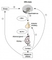
Hypothalamus - Pituitary - Adrenal (HPA)
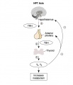
Hypothalamus - Pituitary - Thyroid (HPT)
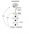
Hypothalamus - Pituitary - Gonad (female) (HPG)
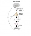
Hypothalamus - Pituitary - Gonad (male) (HPG)


Hypothalamus
- essential for the maintenance of homeostasis.
- regulation of eating, drinking, reproductive and parental behavior, and sleep-wake rhythms.
- controls the autonomic nervous system and hormone secretion.
- rostral-caudal axis - preoptic, anterior, tuberal and mammillary.
- medial-lateral axis - periventricular, medial and lateral zones.

|

|

Human hypothalamus nuclei locations (a - schematic; b- 3D MRI data)
- Pv - paraventricular nucleus
- Sc - suprachiasmatic (ovoid) nucleus
- Links: Hypothalamus Development | Box 7.3 Anatomy of the functional connections between the hypothalamus and pituitary gland
Pituitary


|

|
- hypophysis - pars distalis, pars intermedia, pars nervosa
- pars intermedia - lies between the other 2 parts and consists mainly of colloid filled cysts.
- infundibulum - (infundibular stalk) connection between hypothalamus and the posterior pituitary.
- pars tuberalis - wraps the infundibulum in a highly vascularized sheath, part of the anterior pituitary.
- sits within a cavity in the skull base - the "sella turcica" (Turkish saddle), named historically by its similarity in shape.
- 2 embryonic origins, larger in females than males after pregnancies.
- (phil= likes, phob = hates)
Blood supply
- hypothalamus - blood supply from the circle of Willis.
- pituitary - blood from the inferior (neurohypophysis) and superior (adenohypophysis) hypophyseal arteries.
- inferior hypophyseal artery capillary plexus drains into the dural sinus.
- neural stalk some capillaries (primary capillary network) form about 20+ "short" portal veins that drain into the anterior pituitary gland (secondary capillary network).
- most of anterior lobe has no direct arterial supply, with large fenestrated sinusoidal capillaries.
- hypophyseal vein then drains into systemic venous blood.
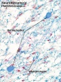
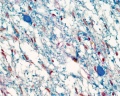
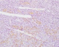
Pituitary - Neurohypophysis
- posterior pituitary
- cells are pituicytes and also present are hypothalamic neurosecretory cell unmyelinated nerve fibres (from the supraoptic and paraventricular nuclei).
- pituicyte - oval or round nuclei are visible, branched cytoplasm, associated with fenestrated capillaries (resemble glial cells).
- Herring bodies are dilations of nerve fibres filled with small neurosecretory vesicles.
- nerve fibres terminate close to capillaries.
- hormones are releasing and release-inhibiting factors produced regulating adenohypophysis activity (as well as 2 other 9 aa hormones).
- Oxytocin - pregnancy stimulates uterine smooth muscle the contraction and lactation milk ejection reflex.
- Antidiuretic hormone - (ADH, vasopressin, arginine vasopressin, AVP) acts on kidneys to concentrate urine (water retention).

|

|
| Neurohypophysis | Herring Bodies (EM) packed with neurosecretory granules. |
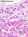
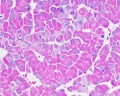
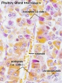
Pituitary - Adenohypophysis
- anterior pituitary - 3 parts pars distalis, pars tuberalis and pars intermedia.
- pars distalis of the adenohypophysis occupies about 75% of the hypophyseal tissue.
- stromal connective tissue - very little visible.
- parenchymal endocrine cells - arranged in irregular clumps or cords between a network of capillaries with large and irregular lumina.
- hormones are proteins or glycoproteins.
- H&E staining identifies 3 cell types:
- acidophil cells (acidophils)
- basophil cells (basophils)
- chromophobe cells

|

|
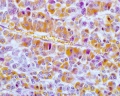
Acidophil cells
- about 65% of all cells.
- rounded and smaller than basophil cells (other stains identify subtypes).
- Somatotrophs - produce growth hormone (GH or somatotropin), stimulates liver cells to produce polypeptide growth factors which stimulate growth (stain with orange G)
- Mammotrophs - (lactotrophs) produce prolactin, maternal numbers increase in third trimester and postnatally in early lactation.
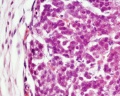
Basophil cells
Based on their hormone products basophils are divided into three subtypes (PAS stain all types reddish).
- Thyrotrophs - produce thyroid stimulating hormone (TSH or thyrotropin).
- Gonadotrophs - produce follicle stimulating hormone (FSH) and luteinizing hormone (LH)
- FSH stimulates in the male seminiferous tubule and female early follicular growth.
- LH stimulates male Leydig cell testosterone production and female oestrogen (estrogen) production, late follicular maturation, formation of corpus luteum.
- Corticotrophs - (or adrenocorticolipotrophs) produce adrenocorticotropic hormone (ACTH or corticotropin) and lipotropin (LPH).
- cell type in the pars intermedia where ACTH and LPH precursor undergoes hydrolysis into melanocyte stimulating hormone (MSH) and other peptides.
Chromophobe cells
- cells are unstained or weakly stained cells.
- either acidophils or basophils in a dormant or recently degranulated stage.
- may also include the secretory stem cells.
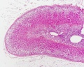
Adrenal


|
Blood supply
|
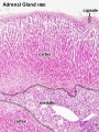
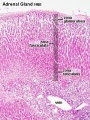
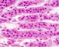
Adrenal Cortex

- divided into three zones (Adrenocorticotropic hormone (ACTH) required for zones 2 and 3)
- zona glomerulosa (about 15%)
- zona fasciculata (about 75%)
- zone reticularis is (about 10%)
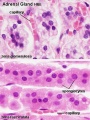
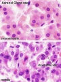
Zona Glomerulosa
- secretes mineralocorticoids.
- Small cells arranged into small rounded groups or curved columns.
- not influenced by ACTH.
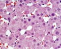
Zona Fasciculata
- secretes glucocorticoids.
- cell cords arranged radially arranged cell cords separated by fenestrated sinusoid capillaries.
- cell nucleus is light and centrally located.
- cell cytoplasm is also light and often "foamy" or "spongy" appearance (due to lipid droplets)
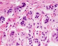
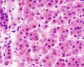
Zona Reticularis
- secretes adrenal androgens.
- cell chords separated by sinusoid spaces.
- small cells with large nucleus, and eosinophilic cytoplasm and less spongy. (lipofucsin accumulates with age, orange colour in H&E).

Zona Glomerulosa and Fasciculata |

Zona Reticularis and Medulla |
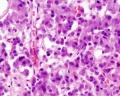
Adrenal Medulla
|

|
Histology Images
Pituitary
Posterior labeled
Posterior unlabelled
Anterior H&E
Anterior H&E
Anterior labeled
PAS/O Overview
Acidophils
Basophils


- Pituitary Histology: Pituitary overview | Anterior H&E | Anterior H&E | Anterior labeled | PAS/O Overview | Acidophils | Basophils | Posterior labeled | Posterior unlabeled | Histology Stains | BGD - Endocrine Histology | Pituitary Development
Adrenal
Cortex and Medulla
Unlabelled Overview
Cortical Zones
Zona Glomerulosa and Fasciculata
Zona Glomerulosa
Zona Fasciculata
Zona Reticularis and Medulla
Zona Reticularis
Medulla
Fetal Cortex
Developing Adult Cortex


- Adrenal Histology: Cortex and Medulla | Unlabelled Overview | Cortical Zones | Zona Glomerulosa and Fasciculata | Zona Glomerulosa | Zona Fasciculata | Zona Reticularis and Medulla | Zona Reticularis | Medulla | Fetal Cortex | Developing Adult Cortex | BGD - Endocrine Histology | Histology Stains | Adrenal Development
Links: Histology | Histology Stains | Blue Histology images copyright Lutz Slomianka 1998-2009. The literary and artistic works on the original Blue Histology website may be reproduced, adapted, published and distributed for non-commercial purposes. See also the page Histology Stains.
Cite this page: Hill, M.A. (2024, April 16) Embryology BGD Lecture - Endocrine Histology. Retrieved from https://embryology.med.unsw.edu.au/embryology/index.php/BGD_Lecture_-_Endocrine_Histology
- © Dr Mark Hill 2024, UNSW Embryology ISBN: 978 0 7334 2609 4 - UNSW CRICOS Provider Code No. 00098G
Additional Information
Not part of today's lecture.
Abnormalities

- Pituitary adenoma - often benign, different effects depending upon cellular function.
- Adrenal tumor - adenoma, neuroblastoma.
Acromegaly PMID 18578866
- acquired disorder related to excessive production of growth hormone (GH)
- characterized by progressive somatic disfigurement (mainly involving the face and extremities) and systemic manifestations.
- prevalence is estimated at 1:140,000-250,000.
- most often diagnosed in middle-aged adults (average age 40 years, men and women equally affected).
- acromegaly is often diagnosed four to more than ten years after its onset.
Cushing's Syndrome PMID 22710101
- (Cushing’s disease, Itsenko-Cushing disease)
- pituitary ACTH dependent Cushing’s syndrome
- increased ACTH from pituitary adenoma.
- rare disease responsible for increased morbidity and mortality.
- Signs and symptoms of hypercortisolism are usually non specific:
- obesity, signs of protein wasting, increased blood pressure, variable levels of hirsutism.
Multiple Endocrine Neoplasia type 1 PMID 17014705
- (MEN1) is a rare autosomal dominant hereditary cancer syndrome
- presented mostly by tumours of the parathyroids, endocrine pancreas and anterior pituitary
- characterised by a very high penetrance and an equal sex distribution.
- occurs in approximately one in 30,000 individuals.
Pheochromocytomas PMID 17156452
- Catecholamine-producing tumors
- may arise in the adrenal medulla (pheochromocytomas) or in extraadrenal chromaffin cells (secreting paragangliomas).
- prevalence is about 0.1% in patients with hypertension and 4% in patients with a fortuitously discovered adrenal mass.
- increase in the production of catecholamines causes symptoms (mainly headaches, palpitations and excess sweating) and signs (mainly hypertension, weight loss and diabetes)
- effects of epinephrine and norepinephrine on α- and β-adrenergic receptors.
History
- 1905 - Introduction of the word "hormone" into the English language. Audio File | Listen Online
- 1930 - First description of the portal system PMID 17104309 PDF
Terms
- acidophilic - (Latin, acidus = sour + Greek, philein = to love) affinity for an acidic dye, such as eosin staining cytoplasmic proteins.
- ACTH - adrenocorticotropic hormone or corticotropin.
- adenoma - a benign tumor of glandular origin.
- adrenaline - (adrenalin, epinephrine) adrenal medulla secreted hormone (also a neurotransmitter), synthesised from tyrosine (amino acid). Many roles throughout the body on many tissues.
- adrenal androgen - zonula reticularis secreted weak steroids or steroid precursors (dehydroepiandrosterone (DHEA), dehydroepiandrosterone sulfate (DHEA-S), and androstenedione).
- basophilic - (Greek, basis = base + philein = to love) affinity for a basic dye, such as haematoxylin, gallocyanin and toluidine blue.
- chromaffin - (Greek, chroma = colour + Latin, affinis = with affinity for) stained with chromium salts; epinephrine-producing cells; para-aortic bodies of Zuckerkandl.
- chromophobe - (Greek, chroma = colour + phobos = fear) cells or granules not taking up any dye readily.
- FSH - follicle stimulating hormone.
- glucocorticoid - adrenal zona fasciculata secretes glucocorticoids (mainly cortisol). Named by their role in glucose metabolism, these steroid hormones bind to the glucocorticoid receptor.
- HPA - Hypothalamic Pituitary Adrenal endocrine axis.
- mineralocorticoid - adrenal zona glomerulosa secreted steroids (mainly aldosterone). Named by their effect on mineral metabolism, acts on the kidney promoting reabsorption of sodium ions (Na+), with water following the salt.
- PAS - Periodic acid-Schiff histological stain.
- TSH - thyroid stimulating hormone or thyrotropin.
See also Histology Glossary
Glossary Links
- Glossary: A | B | C | D | E | F | G | H | I | J | K | L | M | N | O | P | Q | R | S | T | U | V | W | X | Y | Z | Numbers | Symbols | Term Link
Cite this page: Hill, M.A. (2024, April 16) Embryology BGD Lecture - Endocrine Histology. Retrieved from https://embryology.med.unsw.edu.au/embryology/index.php/BGD_Lecture_-_Endocrine_Histology
- © Dr Mark Hill 2024, UNSW Embryology ISBN: 978 0 7334 2609 4 - UNSW CRICOS Provider Code No. 00098G