Cardiovascular System - Circulation Development: Difference between revisions
mNo edit summary |
mNo edit summary |
||
| Line 14: | Line 14: | ||
|-bgcolor="F5FAFF" | |-bgcolor="F5FAFF" | ||
| | | | ||
* '''Infrahepatic inferior caval and azygos vein formation in mammals with different degrees of mesonephric development''' | * '''Infrahepatic inferior caval and azygos vein formation in mammals with different degrees of mesonephric development''' {{#pmid:26659476|PMID26659476}} "The caudal cardinal veins (CCVs) were the only contributors to the inferior caval (IVC) and azygos veins. Development was comparable if temporary vessels that drain the large porcine mesonephros were taken into account. The topography of the CCVs changed concomitant with expansion of adjacent organs (lungs, meso- and metanephroi). The iliac veins arose by gradual extension of the CCVs into the caudal body region. Irrespective of the degree of mesonephric development, the infrarenal part of the IVC developed from the right CCV and the renal part from vascular sprouts of the CCVs in the mesonephros that formed 'subcardinal' veins. The azygos venous system developed from the cranial remnants of the CCVs." | ||
|} | |} | ||
{| class="wikitable mw-collapsible mw-collapsed" | {| class="wikitable mw-collapsible mw-collapsed" | ||
| Line 22: | Line 22: | ||
Search term: [http://www.ncbi.nlm.nih.gov/pubmed/?term=Circulation+Embryology ''Circulation Embryology''] | Search term: [http://www.ncbi.nlm.nih.gov/pubmed/?term=Circulation+Embryology ''Circulation Embryology''] | ||
|} | |} | ||
| Line 40: | Line 38: | ||
Note: Frequently a second renal artery (inferior renal) from abdominal aorta at a lower level, supplies lower portion of kidney | Note: Frequently a second renal artery (inferior renal) from abdominal aorta at a lower level, supplies lower portion of kidney | ||
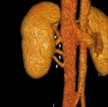
See the review describing the variations in adult renal artery and vein organization. | See the review describing the variations in adult renal artery and vein organization.{{#pmid:20461189|PMID20461189}} of renal vascular anomalies shown in adults using computed tomography. The images below are from that review. | ||
<gallery caption="Renal Arteries"> | <gallery caption="Renal Arteries"> | ||
| Line 53: | Line 51: | ||
====Coronary Arteries Timeline==== | ====Coronary Arteries Timeline==== | ||
Based upon [[Carnegie Collection]] coronary vasculature in 351 staged and serially sectioned human embryos (Carnegie stages 9 to 23). | Based upon [[Carnegie Collection]] coronary vasculature in 351 staged and serially sectioned human embryos (Carnegie stages 9 to 23). {{#pmid:3286038|PMID3286038}} | ||
* [[Carnegie stage 14]] or [[Carnegie stage 15]] - A plexus of blind epicardial capillaries appears on the heart in Carnegie | * [[Carnegie stage 14]] or [[Carnegie stage 15]] - A plexus of blind epicardial capillaries appears on the heart in Carnegie | ||
* [[Carnegie stage 15]], [[Carnegie stage 16]], or [[Carnegie stage 17]] - acquires a coronary sinus connection | * [[Carnegie stage 15]], [[Carnegie stage 16]], or [[Carnegie stage 17]] - acquires a coronary sinus connection | ||
| Line 62: | Line 60: | ||
[[File:Mouse-coronary vessel formation.jpg]] | [[File:Mouse-coronary vessel formation.jpg]] | ||
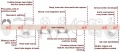
Image showing changes in venous (blue) and arterial (red) marker expression during coronary development; black indicates dedifferentiated venous cells. | Image showing changes in venous (blue) and arterial (red) marker expression during coronary development; black indicates dedifferentiated venous cells.{{#pmid:20336138|PMID20336138}} | ||
:'''Links:''' [[Cardiovascular System - Coronary Circulation Development|Coronary Circulation Development]] | :'''Links:''' [[Cardiovascular System - Coronary Circulation Development|Coronary Circulation Development]] | ||
| Line 71: | Line 69: | ||
===Azygos Vein=== | ===Azygos Vein=== | ||
A recent study, using several species including human, has shown that the caudal cardinal veins are the only contributors to the inferior caval (IVC) and azygos veins. | A recent study, using several species including human, has shown that the caudal cardinal veins are the only contributors to the inferior caval (IVC) and azygos veins.{{#pmid:26659476|PMID26659476}} | ||
Azygos Timeline | Azygos Timeline{{#pmid:25496171|PMID25496171}} | ||
* [[Carnegie stage 11]] to [[Carnegie stage 15]] - caudal cardinal veins extended caudally from the common cardinal vein. | * [[Carnegie stage 11]] to [[Carnegie stage 15]] - caudal cardinal veins extended caudally from the common cardinal vein. | ||
| Line 82: | Line 80: | ||
===Renal Veins=== | ===Renal Veins=== | ||
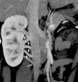
See the review describing the variations in adult renal artery and vein organization | See the review describing the variations in adult renal artery and vein organization{{#pmid:20461189|PMID20461189}} of renal vascular anomalies shown in adults using computed tomography. The images below are from that review. | ||
<gallery caption="Renal Veins"> | <gallery caption="Renal Veins"> | ||
File:Supernumerary_renal_vein_03.jpg|Supernumerary right renal vein | File:Supernumerary_renal_vein_03.jpg|Supernumerary right renal vein | ||
| Line 94: | Line 92: | ||
==Abnormalities== | ==Abnormalities== | ||
* internal carotid artery segmental agenesis - asymptomatic and harmless | * internal carotid artery segmental agenesis - asymptomatic and harmless{{#pmid:27535626|PMID27535626}} | ||
Latest revision as of 11:31, 16 June 2019
| Embryology - 8 Jun 2024 |
|---|
| Google Translate - select your language from the list shown below (this will open a new external page) |
|
العربية | català | 中文 | 中國傳統的 | français | Deutsche | עִברִית | हिंदी | bahasa Indonesia | italiano | 日本語 | 한국어 | မြန်မာ | Pilipino | Polskie | português | ਪੰਜਾਬੀ ਦੇ | Română | русский | Español | Swahili | Svensk | ไทย | Türkçe | اردو | ייִדיש | Tiếng Việt These external translations are automated and may not be accurate. (More? About Translations) |
Introduction

The peripheral circulation, both arterial and venous, are extensively remodelled with embryonic and fetal development. The purpose of this current page is to provide a central resource link to this topic of adult circulatory organization from the embryonic vasculature. Due to the extensive developmental remodelling there are a large number of variations in vascular organization and agenesis.
This general topic is covered in a number of different pages on this site including both coronary circulation and neural circulation.
Some Recent Findings
|
| More recent papers |
|---|
 This table allows an automated computer search of the external PubMed database using the listed "Search term" text link.
More? References | Discussion Page | Journal Searches | 2019 References | 2020 References Search term: Circulation Embryology |
Arteries
Stage 19
 Reconstruction of Carnegie Embryo No. 390 arterial system.
Renal Arteries
- Arise with ascent and inferior branches lost
- Sequential, 25% population have 2 or more renal arteries
- branch of abdominal aorta, divides into 4-5 branches
- each gives off small branches to suprarenal glands, ureter, surrounding cellular tissue and muscles
Note: Frequently a second renal artery (inferior renal) from abdominal aorta at a lower level, supplies lower portion of kidney
See the review describing the variations in adult renal artery and vein organization.[2] of renal vascular anomalies shown in adults using computed tomography. The images below are from that review.
- Renal Arteries
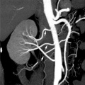
Multiple renal arteries
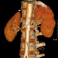
Accessory renal artery


- Links: Renal Vascular Anomalies | Renal
Coronary Arteries
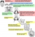
Coronary Arteries Timeline
Based upon Carnegie Collection coronary vasculature in 351 staged and serially sectioned human embryos (Carnegie stages 9 to 23). [3]
- Carnegie stage 14 or Carnegie stage 15 - A plexus of blind epicardial capillaries appears on the heart in Carnegie
- Carnegie stage 15, Carnegie stage 16, or Carnegie stage 17 - acquires a coronary sinus connection
- Carnegie stage 18 - connection of the proximal coronary arteries to the aorta.
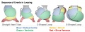
Mouse Coronary Vessels

Image showing changes in venous (blue) and arterial (red) marker expression during coronary development; black indicates dedifferentiated venous cells.[4]
Veins
Azygos Vein
A recent study, using several species including human, has shown that the caudal cardinal veins are the only contributors to the inferior caval (IVC) and azygos veins.[1]
Azygos Timeline[5]
- Carnegie stage 11 to Carnegie stage 15 - caudal cardinal veins extended caudally from the common cardinal vein.
- Carnegie stage 15 to Carnegie stage 18 - caudal cardinal veins sprout ventrally form the sub cardinal vein plexus .
- then caudal part of the left caudal cardinal vein regresses.
- Inferior vena cava - infrarenal part from the right caudal cardinal vein; renal part from subcardinal veins.
- Azygos veins - from the remaining cranial part or sprouting of the caudal cardinal veins.
Renal Veins
See the review describing the variations in adult renal artery and vein organization[2] of renal vascular anomalies shown in adults using computed tomography. The images below are from that review.
- Renal Veins
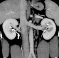
Supernumerary right renal vein
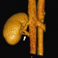
Supernumerary right renal vein
Multiple right renal veins
Multiple right renal veins


- Links: Renal Vascular Anomalies | Renal
Abnormalities
- internal carotid artery segmental agenesis - asymptomatic and harmless[6]
References
- ↑ 1.0 1.1 Hikspoors JP, Mekonen HK, Mommen GM, Cornillie P, Köhler SE & Lamers WH. (2016). Infrahepatic inferior caval and azygos vein formation in mammals with different degrees of mesonephric development. J. Anat. , 228, 495-510. PMID: 26659476 DOI.
- ↑ 2.0 2.1 Kumar S, Neyaz Z & Gupta A. (2010). The utility of 64 channel multidetector CT angiography for evaluating the renal vascular anatomy and possible variations: a pictorial essay. Korean J Radiol , 11, 346-54. PMID: 20461189 DOI.
- ↑ Hutchins GM, Kessler-Hanna A & Moore GW. (1988). Development of the coronary arteries in the embryonic human heart. Circulation , 77, 1250-7. PMID: 3286038
- ↑ Red-Horse K, Ueno H, Weissman IL & Krasnow MA. (2010). Coronary arteries form by developmental reprogramming of venous cells. Nature , 464, 549-53. PMID: 20336138 DOI.
- ↑ Hikspoors JP, Soffers JH, Mekonen HK, Cornillie P, Köhler SE & Lamers WH. (2015). Development of the human infrahepatic inferior caval and azygos venous systems. J. Anat. , 226, 113-25. PMID: 25496171 DOI.
- ↑ Alexandre AM, Visconti E, Schiarelli C, Frassanito P & Pedicelli A. (2016). Bilateral Internal Carotid Artery Segmental Agenesis: Embryology, Common Collateral Pathways, Clinical Presentation, and Clinical Importance of a Rare Condition. World Neurosurg , 95, 620.e9-620.e15. PMID: 27535626 DOI.
Reviews
Articles
Search Pubmed
Search May 2010
- Cardiovascular System Development All (63457) Review (10735) Free Full Text (15717)
Search Pubmed: Coronary Circulation Development
Additional Images
See also Category:Heart ILP and Category:Heart
Historic image
Heart Development Timeline
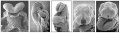
Human heart SEM
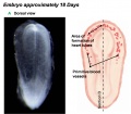
Early Heart Tube (Dorsal)
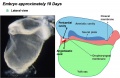
Early Heart Tube (Lateral)
Heart Tube Segments
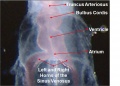
Heart Looping Sequence
Molecular & Genetic Cardiac Development Factors
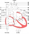
Adult heart blood flow cartoon


.jpg)
.jpg)


External Links
External Links Notice - The dynamic nature of the internet may mean that some of these listed links may no longer function. If the link no longer works search the web with the link text or name. Links to any external commercial sites are provided for information purposes only and should never be considered an endorsement. UNSW Embryology is provided as an educational resource with no clinical information or commercial affiliation.
Glossary Links
- Glossary: A | B | C | D | E | F | G | H | I | J | K | L | M | N | O | P | Q | R | S | T | U | V | W | X | Y | Z | Numbers | Symbols | Term Link
Cite this page: Hill, M.A. (2024, June 8) Embryology Cardiovascular System - Circulation Development. Retrieved from https://embryology.med.unsw.edu.au/embryology/index.php/Cardiovascular_System_-_Circulation_Development
- © Dr Mark Hill 2024, UNSW Embryology ISBN: 978 0 7334 2609 4 - UNSW CRICOS Provider Code No. 00098G