File:Blood capillary EM 01.jpg: Difference between revisions
From Embryology
No edit summary |
|||
| Line 10: | Line 10: | ||
===Reference=== | ===Reference=== | ||
<pubmed>21702933</pubmed>| [http://www.ncbi.nlm.nih.gov/pmc/articles/PMC3141733 PMC3141733] | [http://www.biomedcentral.com/1471-2121/12/29 BMC Cell Biol.] | |||
Original file name: 1471-2121-12-29-2.jpg | Original file name: 1471-2121-12-29-2.jpg | ||
[[Category:Blood Vessel]] [[Category:Cardiovascular]] [[Category:Electron Micrograph]] | [[Category:Blood Vessel]] [[Category:Cardiovascular]] [[Category:Electron Micrograph]] | ||
Revision as of 10:28, 5 February 2012
Blood Capillary Electron Micrograph
A blood capillary lumen (lu) lined by an endothelial cell (en) is surrounded by a continuous basal lamina (arrowhead) in which is incorporated a pericyte (p).
Extracellular matrix collagen bundles at top of image.
Scale bar 1 μm
Panel D cropped from Figure 2. (1471-2121-12-29-2.jpg) Contrast and size adjusted.
Reference
<pubmed>21702933</pubmed>| PMC3141733 | BMC Cell Biol.
Original file name: 1471-2121-12-29-2.jpg
File history
Click on a date/time to view the file as it appeared at that time.
| Date/Time | Thumbnail | Dimensions | User | Comment | |
|---|---|---|---|---|---|
| current | 10:25, 5 February 2012 | 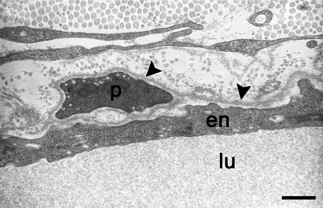 | 1,107 × 714 (260 KB) | S8600021 (talk | contribs) | ==Blood Capillary Electron Micrograph== A blood capillary lined by an endothelial cell (en) is surrounded by a continuous basal lamina (arrowhead) in which is incorporated a pericyte (p) Panel D cropped from Figure 2. (1471-2121-12-29-2.jpg) Contrast an |
You cannot overwrite this file.
File usage
The following 2 pages use this file: