Palate Development: Difference between revisions
mNo edit summary |
mNo edit summary |
||
| (44 intermediate revisions by the same user not shown) | |||
| Line 2: | Line 2: | ||
== Introduction == | == Introduction == | ||
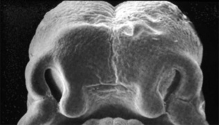
[[File:Stage18 em11.jpg|thumb|300px|Human Embryo Face ([[Week 7]], [[Carnegie stage 18]], 44 - 48 days, CRL 13 - 17 mm)]] | [[File:Stage18 em11.jpg|thumb|300px|Human Embryo Face ([[Week 7]], [[Carnegie stage 18]], 44 - 48 days, CRL 13 - 17 mm)]] | ||
The palate has two | The palate anatomically separates the nasal cavity from the oral cavity and structurally has a bony (hard) anterior component and a muscular (soft) posterior component ending with the uvula. The oral side of the palate is covered with a squamous stratified (pluristratified) epithelium. The surface of the hard palate of most mammalian species is further thrown into a series of transversal palatal ridges or ''rugae palatinae''. Both the palatal ridge number and arrangement are also species specific. | ||
Neural crest has a major contribution to the palate development and there are a number of molecular, mechanical and morphological steps in involving the fusion of contributing structures including a key epithelial to mesenchymal transition. In palate formation there are two main and separate times and events of development, during embryonic (primary palate) and an early fetal (secondary palate). This separation of events into embryonic and fetal period corresponds closely to the classification of associated palate abnormalities. | |||
| Line 11: | Line 14: | ||
The secondary palate can also be divided in two anatomical parts: | The secondary palate can also be divided in two anatomical parts: | ||
# anterior hard palate - ossified (contributions from the maxilla and palatine bones) | # anterior hard palate - ossified (contributions from the maxilla and palatine bones). | ||
# posterior soft palate - muscular. | # posterior soft palate - muscular. | ||
{{Palate Links}} | |||
<br> | |||
{{Head Links}} | {{Head Links}} | ||
| Line 23: | Line 25: | ||
== Some Recent Findings == | == Some Recent Findings == | ||
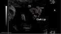
[[File:Cleft_lip_01.jpg|thumb|Ultrasound - Cleft Lip]] | [[File:Cleft_lip_01.jpg|thumb|Ultrasound - Cleft Lip]] | ||
[[File:Pharyngeal_arch_cartilages.jpg|thumb]] | |||
{| | {| | ||
|-bgcolor="F5FAFF" | |-bgcolor="F5FAFF" | ||
| | | | ||
* ''' | * '''Short stature homeobox 2 (SHOX2) regulates osteogenic differentiation and pattern formation during hard palate development in mice'''{{#pmid:31649032|PMID31649032}} "During mammalian palatogenesis, cranial neural crest-derived mesenchymal cells undergo osteogenic differentiation and form the hard palate, which is divided into palatine process of the maxilla and the palatine. However, it remains unknown whether these bony structures originate from the same cell lineage and how the hard palate is patterned at the molecular level. Using mice, here we report that deficiency in short stature homeobox 2 (Shox2), a transcriptional regulator whose expression is restricted to the anterior palatal mesenchyme, leads to a defective palatine process of the maxilla, but does not affect the palatine. Shox2 overexpression in palatal mesenchyme resulted in a hyperplastic palatine process of the maxilla and a hypoplastic palatine. RNA-Seq and assay for transposase-accessible chromatin (ATAC)-Seq analyses revealed that SHOX2 controls the expression of pattern-specification and skeletogenic genes associated with accessible chromatin in the anterior palate. This highlighted a lineage-autonomous function of SHOX2 in patterning and osteogenesis of the hard palate. H3K27ac ChIP-Seq and transient transgenic enhancer assays revealed that SHOX2 binds distal-acting cis-regulatory elements in an anterior palate-specific manner. Our results suggest that the palatine process of the maxilla and palatine arise from different cell lineages and differ in ossification mechanisms. SHOX2 evidently controls osteogenesis of a cell lineage and contributes to the palatine process of the maxilla by interacting with distal cis-regulatory elements to regulate skeletogenic gene expression and to pattern the hard palate. Genome-wide SHOX2 occupancy in the developing palate may provide a marker for identifying active anterior palate-specific gene enhancers." | ||
* ''' | |||
* ''' | * '''{{TGF-beta}} (TGF-β) Signaling and the Epithelial-Mesenchymal Transition during Palatal Fusion'''{{#pmid:30463190|PMID30463190}} "Signaling by transforming growth factor {{TGF-beta}} plays an important role in development, including in palatogenesis. The dynamic morphological process of palatal fusion occurs to achieve separation of the nasal and oral cavities. Critically and specifically important in palatal fusion are the medial edge epithelial (MEE) cells, which are initially present at the palatal midline seam and over the course of the palate fusion process are lost from the seam, due to cell migration, {{epithelial mesenchymal transition}} (EMT), and/or programed cell death. ...{{mouse}} TGF-β is highly regulated both temporally and spatially, with TGF-β3 and Smad2 being the preferentially expressed signaling molecules in the critical cells of the fusion processes." | ||
* ''' | |||
* ''' | * '''Development of Hard and Soft Palate During the Fetal Period and Hard Palate Asymmetry'''{{#pmid:30320695|PMID30320695}} "In the present study, it was aimed to perform the morphometric analysis of the hard and soft palate in fetal cadavers and evaluate hard palate asymmetry during the fetal development. The development of the palate was investigated in 40 (21 males, 19 females) fetal materials aged between the 17th and 40th gestational week. In this study, distances between the incisive papilla-staurion (Ip-Sr), staurion-posterior nasal spine (Sr-Pns), incisive papilla-greater palatine foramen (Ip-Gpf) on the right and left sides, Sr-Gpf, and Pns-Gpf were measured. In cases with asymmetry, the ratio of asymmetry was determined in percentage using the asymmetry index. Moreover, angular values between Ip-Sr-Gpf and Ip-Pns-Gpf on the right and left sides were measured, and the right and left side values were compared with each other." | ||
* '''Ectopic Hedgehog Signaling Causes Cleft Palate and Defective Osteogenesis'''{{#pmid:29975848|PMID29975848}} "Cleft palate is a common birth defect that frequently occurs in human congenital malformations caused by mutations in components of the Sonic Hedgehog ({{SHH}}) signaling cascade. Shh is expressed in dynamic, spatiotemporal domains within epithelial rugae and plays a key role in driving epithelial-mesenchymal interactions that are central to development of the secondary palate. However, the gene regulatory networks downstream of Hedgehog (Hh) signaling are incompletely characterized. Here, we show that ectopic Hh signaling in the palatal mesenchyme disrupts oral-nasal patterning of the neural crest cell-derived ectomesenchyme of the palatal shelves, leading to defective palatine bone formation and fully penetrant cleft palate. We show that a series of Fox transcription factors, including the novel direct target Foxl1, function downstream of Hh signaling in the secondary palate. Furthermore, we demonstrate that {{Wnt}}/bone morphogenetic protein ({{BMP}}) antagonists, in particular Sostdc1, are positively regulated by Hh signaling, concomitant with down-regulation of key regulators of osteogenesis and BMP signaling effectors." | |||
* '''Three-dimensional imaging of palatal muscles in the human embryo and fetus: Development of levator veli palatini and clinical importance of the lesser palatine nerve'''{{#pmid:26509917|PMID26509917}} "The development of LVP in human embryos and fetuses has not been systematically analyzed using the Carnegie stage (CS) to standardize documentation of development. The anlage of LVP starts to develop at CS {{CS21}} beneath the aperture of the auditory tube (AT) to the pharynx. At CS {{CS23}}, LVP runs along AT over its full length, as evidenced by three-dimensional image reconstruction. In the {{fetal}} stage, the lesser palatine nerve (LPN) is in contact with LVP. The positional relationship between LVP and AT three-dimensionally, suggesting that LVP might be derived from the second branchial arch. Based on histological evidence, we hypothesize that motor components from the facial nerve may run along LPN, believed to be purely sensory. The multiple innervation of LVP by LPN and pharyngeal plexus may explain the postpalatoplasty discrepancy between the partial impairment in articulation vs. the functional restoration of deglutition. That is, the contribution of LPN is greater in articulation than in deglutition." | |||
* '''Molecular Anatomy of Palate Development'''{{#pmid:26168040|PMID26168040}} "The [https://www.facebase.org NIH FACEBASE] consortium was established in part to create a central resource for craniofacial researchers. One purpose is to provide a molecular anatomy of craniofacial development. To this end we have used a combination of laser capture microdissection and RNA-Seq to define the gene expression programs driving development of the murine palate. We focused on the {{ME14.5}} palate, soon after medial fusion of the two palatal shelves. The palate was divided into multiple compartments, including both medial and lateral, as well as oral and nasal, for both the anterior and posterior domains. A total of 25 RNA-Seq datasets were generated. The results provide a comprehensive view of the region specific expression of all transcription factors, growth factors and receptors. Paracrine interactions can be inferred from flanking compartment growth factor/receptor expression patterns. The results are validated primarily through very high concordance with extensive previously published gene expression data for the developing palate." | |||
|} | |} | ||
{| class="wikitable mw-collapsible mw-collapsed" | |||
! More recent papers | |||
|- | |||
| [[File:Mark_Hill.jpg|90px|left]] {{Most_Recent_Refs}} | |||
{| class="wikitable collapsible collapsed" | Search term: [http://www.ncbi.nlm.nih.gov/pubmed/?term=Palate+Development ''Palate Development''] | [http://www.ncbi.nlm.nih.gov/pubmed/?term=Palate+Embryology ''Palate Embryology''] | [http://www.ncbi.nlm.nih.gov/pubmed/?term=Embryonic+Palate ''Embryonic Palate''] | [http://www.ncbi.nlm.nih.gov/pubmed/?term=Fetal+Palate ''Fetal Palate''] | [http://www.ncbi.nlm.nih.gov/pubmed/?term=Cleft+Palate ''Cleft Palate''] | [http://www.ncbi.nlm.nih.gov/pubmed/?term=levator+veli+palatini+Development levator veli palatini Development] | ||
! | |||
|} | |||
{| class="wikitable mw-collapsible mw-collapsed" | |||
! Older papers | |||
|- | |- | ||
| | | {{Older papers}} | ||
* '''Regulation of the Epithelial Adhesion Molecule CEACAM1 Is Important for Palate Formation'''{{#pmid:23613893|PMID23613893}} "Cleft palate results from a mixture of genetic and environmental factors and occurs when the bilateral palatal shelves fail to fuse. The objective of this study was to search for new genes involved in mouse palate formation. Gene expression of murine embryonic palatal tissue was analyzed at various developmental stages before, during, and after palate fusion using GeneChip® microarrays. Ceacam1 was one of the highly up-regulated genes during palate formation, and this was confirmed by quantitative real-time PCR. Immunohistochemical staining showed that CEACAM1 was present in prefusion palatal epithelium and was degraded during fusion. ...These results suggest that CEACAM1 has roles in the initiation of palatal fusion via epithelial cell adhesion." | |||
* '''Role of GSK-3β in the Osteogenic Differentiation of Palatal Mesenchyme'''{{#pmid:22022457|PMID22022457}} "Here, we identify a critical role for GSK-3β in palatogenesis through its direct regulation of canonical Wnt signaling. These findings shed light on critical developmental pathways involved in palatogenesis and may lead to novel molecular targets to prevent cleft palate formation." | |||
* '''Ephrin reverse signaling controls palate fusion via a PI3 kinase-dependent mechanism'''{{#pmid:21246652|PMID21246652}} "Secondary palate fusion requires adhesion and epithelial-to-mesenchymal transition (EMT) of the epithelial layers on opposing palatal shelves. This EMT requires transforming growth factor β3 (TGFβ3), and its failure results in cleft palate. Ephrins, and their receptors, the Ephs, are responsible for migration, adhesion, and midline closure events throughout development. Ephrins can also act as signal-transducing receptors in these processes, with the Ephs serving as ligands (termed "reverse" signaling). We found that activation of ephrin reverse signaling in chicken palates induced fusion in the absence of TGFβ3, and that PI3K inhibition abrogated this effect. Further, blockage of reverse signaling inhibited TGFβ3-induced fusion in the chicken and natural fusion in the mouse. Thus, ephrin reverse signaling is necessary and sufficient to induce palate fusion independent of TGFβ3." | |||
* '''A genome-wide association study of cleft lip with and without cleft palate identifies risk variants near MAFB and ABCA4'''{{#pmid:20436469|PMID 20436469}} "Case-parent trios were used in a genome-wide association study of cleft lip with and without cleft palate. SNPs near two genes not previously associated with cleft lip with and without cleft palate (MAFB, most significant SNP rs13041247, with odds ratio (OR) per minor allele = 0.704, 95% CI 0.635-0.778, P = 1.44 x 10(-11); and ABCA4, most significant SNP rs560426, with OR = 1.432, 95% CI 1.292-1.587, P = 5.01 x 10(-12)) and two previously identified regions (at chromosome {{Chr8}}q24 and IRF6) attained genome-wide significance." | |||
* '''A dosage-dependent role for Spry2 in growth and patterning during palate development'''{{#pmid:17693063|PMNID17693063}} "The formation of the palate involves the coordinated outgrowth, elevation and midline fusion of bilateral shelves leading to the separation of the oral and nasal cavities. Reciprocal signaling between adjacent fields of epithelial and mesenchymal cells directs palatal shelf growth and morphogenesis. Loss of function mutations in genes encoding FGF ligands and receptors have demonstrated a critical role for FGF signaling in mediating these epithelial-mesenchymal interactions. The Sprouty family of genes encode modulators of FGF signaling. We have established that mice carrying a deletion that removes the FGF signaling antagonist Spry2 have cleft palate." | |||
|} | |} | ||
== Textbooks == | == Textbooks == | ||
* '''The Developing Human: Clinically Oriented Embryology''' (8th Edition) by Keith L. Moore and T.V.N Persaud - Moore & Persaud Chapter Chapter 10 The Pharyngeal Apparatus pp201 - 240. | * '''The Developing Human: Clinically Oriented Embryology''' (8th Edition) by Keith L. Moore and T.V.N Persaud - Moore & Persaud Chapter Chapter 10 The Pharyngeal Apparatus pp201 - 240. | ||
* '''Larsen’s Human Embryology''' by GC. Schoenwolf, SB. Bleyl, PR. Brauer and PH. Francis-West - Chapter 12 Development of the Head, the Neck, the Eyes, and the Ears pp349 - 418. | * '''Larsen’s Human Embryology''' by GC. Schoenwolf, SB. Bleyl, PR. Brauer and PH. Francis-West - Chapter 12 Development of the Head, the Neck, the Eyes, and the Ears pp349 - 418. | ||
| Line 63: | Line 80: | ||
:'''Links:''' [[Movies]] | | :'''Links:''' [[Movies]] | {{ultrasound}} | ||
== Development Overview == | == Development Overview == | ||
| Line 71: | Line 88: | ||
* '''week 9''' - secondary palate shelves fuse, separating oral and nasal cavities. | * '''week 9''' - secondary palate shelves fuse, separating oral and nasal cavities. | ||
Embryonic Period | ===Embryonic Period=== | ||
* (week 4) - pharyngeal arch formation in rostrocaudal sequence (1, 2, 3, 4 and 6) | * (week 4) - pharyngeal arch formation in rostrocaudal sequence (1, 2, 3, 4 and 6) | ||
* First pharyngeal arch - upper maxillary (pair) and lower mandibular prominences | * First pharyngeal arch - upper maxillary (pair) and lower mandibular prominences | ||
* Late embryonic period - maxillary prominences fuse with frontonasal prominence forming upper jaw (maxilla and upper lip) | * Late embryonic period - maxillary prominences fuse with frontonasal prominence forming upper jaw (maxilla and upper lip) | ||
<gallery> | <gallery mode="packed-hover" caption="Embryonic Palate"> | ||
File:Stage16 em01.jpg|Week 5 [[ | File:Stage16 em01.jpg|Week 5 [[Carnegie stage 16|stage 16]] | ||
File:Stage17 em01.jpg|Week 6 [[ | File:Stage17 em01.jpg|Week 6 [[Carnegie stage 17|stage 17]] | ||
File:Stage18 em01.jpg|Week 6.5 [[ | File:Stage18 em01.jpg|Week 6.5 [[Carnegie stage 18|stage 18]] | ||
File: | File:Stage19 em01.jpg|Week 7 [[Carnegie stage 19|stage 19]] | ||
File:Stage_22_image_061.jpg|Week 8 [[Carnegie stage 22|stage 22]] | |||
File:Stage23 embryo oral cavity 03.jpg|Week 8 [[Carnegie stage 23|stage 23]] | |||
File:Stage23 embryo oral cavity 04.jpg|Week 8 [[Carnegie stage 23|stage 23]] | |||
</gallery> | </gallery> | ||
Fetal Period | |||
===Fetal Period=== | |||
{| | {| | ||
| | | | ||
| Line 100: | Line 121: | ||
* branchial arch (Gk. branchia= gill) | * branchial arch (Gk. branchia= gill) | ||
* arch consists of all 3 trilaminar embryo layers | * arch consists of all 3 trilaminar embryo layers | ||
* ectoderm- outside | * ectoderm - outside | ||
* mesoderm- core of mesenchyme | * mesoderm - core of mesenchyme | ||
* endoderm- inside | * endoderm - inside | ||
===Neural Crest === | ===Neural Crest === | ||
* Mesenchyme invaded by neural crest generating connective tissue components | * Mesenchyme invaded by {{neural crest}} generating connective tissue components | ||
* cartilage, bone, ligaments | * cartilage, bone, ligaments | ||
* arises from midbrain and hindbrain region | * arises from midbrain and hindbrain region | ||
| Line 120: | Line 141: | ||
===Frontonasal Process=== | ===Frontonasal Process=== | ||
The frontonasal process (FNP) forms the majority of the superior part of the early face primordia. It later fuses with the maxillary component of the first pharyngeal arch to form the upper jaw. Failure of this fusion event during the embryonic period leads to cleft lip. Under the surface ectoderm the process mesenchyme consists of two cell populations; neural crest cells, forming the connective tissues; and the mesoderm forming the endothelium of the vascular network. | The frontonasal process (FNP) forms the majority of the superior part of the early face primordia. It later fuses with the maxillary component of the first pharyngeal arch to form the upper jaw. Failure of this fusion event during the embryonic period leads to cleft lip. Under the surface ectoderm the process mesenchyme consists of two cell populations; neural crest cells, forming the connective tissues; and the mesoderm forming the endothelium of the vascular network. | ||
A chicken developmental model study has identified a specific surface region, the Frontonasal Ectodermal Zone (FEZ), initially induced by bone morphogenetic proteins that appears to regulate the future growth and patterning of the frontonasal process. The specific frontonasal ectodermal zone was located in the frontonasal process ectoderm flanking a boundary between Sonic hedgehog ( | A chicken developmental model study has identified a specific surface region, the Frontonasal Ectodermal Zone (FEZ), initially induced by bone morphogenetic proteins that appears to regulate the future growth and patterning of the frontonasal process. The specific frontonasal ectodermal zone was located in the frontonasal process ectoderm flanking a boundary between Sonic hedgehog ({{SHH}}) and Fibroblast growth factor 8 ({{FGF}}8) expression domains.{{#pmid:18028903|PMID18028903}} | ||
== Embryonic Palate== | == Embryonic Palate== | ||
| Line 130: | Line 149: | ||
| | | | ||
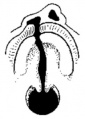
Human primary palate | Human primary palate | ||
* develops between embryonic stages | * develops between embryonic stages {{CS15}} and {{CS18}}.{{#pmid:8227288|PMID8227288}} | ||
* fusion in the human embryo between stage | * fusion in the human embryo between stage {{CS17}} and {{CS18}}, from an epithelial seam to the mesenchymal bridge. | ||
| [[File:Stage17-18 Primary palate.gif]] | | [[File:Stage17-18 Primary palate.gif]] | ||
| Line 137: | Line 156: | ||
<gallery> | <gallery> | ||
File:Stage16 em01.jpg|Week 5 [[:File:Stage16 em01.jpg|Stage | File:Stage16 em01.jpg|Week 5 [[:File:Stage16 em01.jpg|Stage {{CS16}}]] | ||
File:Stage17 em01.jpg|Week 6 [[:File:Stage17 em01.jpg|Stage | File:Stage17 em01.jpg|Week 6 [[:File:Stage17 em01.jpg|Stage {{CS17}}]] | ||
File:Stage18 em01.jpg|Week 6.5 [[:File:Stage18 em01.jpg|Stage | File:Stage18 em01.jpg|Week 6.5 [[:File:Stage18 em01.jpg|Stage {{CS18}}]] | ||
File:Stage19 em01.jpg|Week 7 [[:File:Stage19 em01.jpg|Stage | File:Stage19 em01.jpg|Week 7 [[:File:Stage19 em01.jpg|Stage {{CS19}}]] | ||
</gallery> | </gallery> | ||
:'''EM Links:''' [[:File:Stage16 em01.jpg|Image - stage 16]] | [[:File:Stage17 em01.jpg|Image - stage 17]] | [[:File:Stage18 em01.jpg|Image - stage 18]] | [[:File:Stage19 em01.jpg|Image - stage 19 | :'''EM Links:''' [[:File:Stage16 em01.jpg|Image - stage 16]] | [[:File:Stage17 em01.jpg|Image - stage 17]] | [[:File:Stage18 em01.jpg|Image - stage 18]] | [[:File:Stage19 em01.jpg|Image - stage 19]] | ||
==Fetal Palate== | ==Fetal Palate== | ||
{| | |||
| valign=top|Secondary palate, fusion in the human embryo in [[Week 9|week 9]]. This requires the early palatal shelves growth, elevation, and fusion. There are many fusion events occurring during this period between each palatal shelf, to the primary palate, and also to the nasal septum. | |||
[[:File:Palatal shelves animation.gif|palatal shelf elevation]] | [[:File:palate.gif|secondary palate]] | |||
<gallery> | <gallery> | ||
File:Fetal week 10 hard palate 04.jpg|Hard and soft palate | File:Fetal week 10 hard palate 04.jpg|Hard and soft palate | ||
| Line 158: | Line 180: | ||
File:Fetal week 10 hard palate 02.jpg|Detail - hard palate seam | File:Fetal week 10 hard palate 02.jpg|Detail - hard palate seam | ||
</gallery> | </gallery> | ||
| valign=top|[[File:Fetal palate growth graph.jpg|alt=Fetal palate growth graph.|500px]] | |||
Fetal Palate Growth (second trimester){{#pmid:14245486|PMID14245486}} | |||
|- | |||
| [[File:Fetal week 9 hard palate fusion 01.jpg|400px]] | |||
[[File:Bailey141.jpg|400px]] | Fetal week 9 ({{GA}} week 11) hard palate fusion | ||
| [[File:Bailey141.jpg|400px]] | |||
Ventral aspect of hard palate of human embryo of 80 mm | Ventral aspect of hard palate of human embryo of 80 mm | ||
|} | |||
==Soft Palate== | |||
The soft palate mechanism of closure has not yet been determined, with several existing theories. A recent study{{#pmid:26299693|PMID26299693}} of embryos from the late embryonic-early fetal period (54 to 74 days post-conception) has identified the timing of soft palate closure. | |||
* '''57 days''' - Late embryonic (Carnegie stage {{CS23}}), epithelial seam present throughout the soft palate | |||
* '''64 days''' - Early fetal ([[Week 9]]), epithelium only persists in the most posterior regions of the soft palate | |||
==Postnatal Head Changes== | |||
Head Growth continues postnatally with fontanelle initially allowing head distortion on birth and early growth. These bony plates remain unfused to allow growth, puberty growth of face. | |||
The palate also grows postnatally through childhood and becomes more elevated (arched) forming the "palatine vault", with different (but insignificant) growth between the genders.{{#pmid:23314328|PMID23314328}} | |||
:'''Links:''' [[Postnatal Development]] | |||
==Animal Palate== | ==Animal Palate== | ||
| Line 180: | Line 220: | ||
[[File:Mouse_palate_gene_expression_01.jpg|600px]] | [[File:Mouse_palate_gene_expression_01.jpg|600px]] | ||
Mouse (E13.5) Palatal Shelf Wnt5a, Osr2 and Pax9 Expression. | Mouse (E13.5) Palatal Shelf Wnt5a, Osr2 and Pax9 Expression.{{#pmid:24433583|PMID24433583}} | ||
<gallery> | <gallery> | ||
| Line 214: | Line 254: | ||
:'''Links:''' [[Developmental_Signals_-_Bone_Morphogenetic_Protein|Bone Morphogenetic Protein]] | :'''Links:''' [[Developmental_Signals_-_Bone_Morphogenetic_Protein|Bone Morphogenetic Protein]] | ||
== Abnormalities== | == Abnormalities== | ||
Note there are specific pages for both [[Abnormal_Development_-_Cleft_Lip_and_Palate|Cleft Lip and Palate]] and [[Abnormal Development - Cleft Palate|Cleft Palate]]. | |||
===Clinical Images=== | |||
'''Cleft Lip/Palate''' | |||
<gallery> | |||
File:Cleft_lip_001.jpg|Unilateral cleft lip | |||
File:Cleft_lip_002.jpg|Unilateral cleft lip | |||
File:Cleft_lip_003.jpg|Complete Unilateral cleft lip and palate | |||
File:Cleft_lip_004.jpg|Complete Unilateral cleft lip and palate | |||
File:Cleft_lip_005.jpg|Complete bilateral cleft lip and palate | |||
File:Cleft_lip_006.jpg|Complete bilateral cleft lip and palate | |||
File:Cleft_lip_007.jpg|Cheiloplasty - surgical repair of the lip | |||
</gallery> | |||
'''Cleft Palate''' | |||
<gallery> | |||
File:Cleft_palate_001.jpg|Incomplete cleft palate | |||
File:Cleft_palate_002.jpg|Cleft palate | |||
File:Cleft_palate_003.jpg|Palatoplasty - surgical repair of the palate | |||
</gallery> | |||
[[File:Australian abnormalities 81-92 git.jpg|thumb|350px|Cleft Palate - Australia (1981-1992)<ref>P. Lancaster and E. Pedisich, '''Congenital Malformations Australia''' 1981-1992, ISSN 1321-835.</ref>]] | [[File:Australian abnormalities 81-92 git.jpg|thumb|350px|Cleft Palate - Australia (1981-1992)<ref>P. Lancaster and E. Pedisich, '''Congenital Malformations Australia''' 1981-1992, ISSN 1321-835.</ref>]] | ||
The way in which the upper jaw forms from fusion of the smaller upper prominence of the first pharyngeal arch leads to a common congenital defect in this region called "clefting", which may involve either the upper lip, the palate or both structures. | The way in which the upper jaw forms from fusion of the smaller upper prominence of the first pharyngeal arch leads to a common congenital defect in this region called "clefting", which may involve either the upper lip, the palate or both structures. | ||
{{Australian Palate abnormalities 2002-2003}} | {{Australian Palate abnormalities 2002-2003}} | ||
{{Global Orofacial Cleft Rate (1950 - 2015)}} | |||
===International Classification of Diseases - Cleft Palate=== | ===International Classification of Diseases - Cleft Palate=== | ||
| Line 274: | Line 340: | ||
=== Cleft Lip === | === Cleft Lip === | ||
{| | {| | ||
| [[File:Bilateral_cleft_palate.jpg|200px|Bilateral cleft palate]] | | [[File:Bilateral_cleft_palate.jpg|200px|Bilateral cleft palate]] | ||
| Line 283: | Line 350: | ||
|} | |} | ||
'''Midline Cleft Lip Genes''' | {| class="wikitable collapsible collapsed" | ||
! Cleft Lip Genes{{#pmid:21331089|PMID21331089}} | |||
|- | |||
| '''Midline Cleft Lip Genes''' | |||
* Opitz G/BBB (MID1) | * Opitz G/BBB (MID1) | ||
* Oro-facial-digital type I (OFD1) | * Oro-facial-digital type I (OFD1) | ||
|- | |||
'''Cleft Lip (+/− cleft palate) Genes''' | | | ||
'''Cleft Lip (+/− cleft palate) Genes''' | |||
{| | {| | ||
! Syndrome | ! Syndrome | ||
| Line 346: | Line 417: | ||
| WNT3 | | WNT3 | ||
|} | |} | ||
|} | |||
=== Cleft Palate === | === Cleft Palate === | ||
[[File:Cleft_palate_02.jpg|thumb|An 8 month old infant with an extensive cleft palate associated with Bamforth- Lazarus syndrome. | [[File:Cleft_palate_02.jpg|thumb|An 8 month old infant with an extensive cleft palate associated with Bamforth- Lazarus syndrome.{{#pmid:20537182|PMID20537182}}]] | ||
{| | {| | ||
| [[File:cleft_palate.jpg|200px|Cleft palate]] | | [[File:cleft_palate.jpg|200px|Cleft palate]] | ||
| Line 360: | Line 431: | ||
{| class="wikitable collapsible collapsed" | {| class="wikitable collapsible collapsed" | ||
! Cleft Palate Only Genes | ! Cleft Palate Only Genes{{#pmid:21331089|PMID21331089}} | ||
|- | |- | ||
| | | | ||
| Line 468: | Line 539: | ||
|} | |} | ||
'''Links:''' | |||
'''Links:''' [http://www.ncbi.nlm.nih.gov/omim/119530 OMIM Orofacial Cleft with or without cleft palate] | |||
'''Search Pubmed Now:''' [http://www.ncbi.nlm.nih.gov/entrez/query.fcgi?CMD=search&DB=pubmed&term=cleft%20lip cleft lip] | [http://www.ncbi.nlm.nih.gov/entrez/query.fcgi?CMD=search&DB=pubmed&term=cleft%20palate cleft palate] | '''Search Pubmed Now:''' [http://www.ncbi.nlm.nih.gov/entrez/query.fcgi?CMD=search&DB=pubmed&term=cleft%20lip cleft lip] | [http://www.ncbi.nlm.nih.gov/entrez/query.fcgi?CMD=search&DB=pubmed&term=cleft%20palate cleft palate] | ||
| Line 488: | Line 564: | ||
===Folate=== | ===Folate=== | ||
A recent study of periconceptional folate supplementation using the Cochrane Pregnancy and Childbirth Group's Trials Register (July 2010) identified no statistically significant evidence of any effects on prevention of cleft palate and cleft lip at birth. | A recent study of periconceptional folate supplementation using the Cochrane Pregnancy and Childbirth Group's Trials Register (July 2010) identified no statistically significant evidence of any effects on prevention of cleft palate and cleft lip at birth.{{#pmid:20927767|PMID20927767}} | ||
== References == | == References == | ||
| Line 500: | Line 576: | ||
===Reviews=== | ===Reviews=== | ||
Indian J Plast Surg. 2009 October; 42(Suppl):[http://www.ncbi.nlm.nih.gov/pmc/issues/185089 Cleft Lip and Palate Issue] | Indian J Plast Surg. 2009 October; 42(Suppl):[http://www.ncbi.nlm.nih.gov/pmc/issues/185089 Cleft Lip and Palate Issue] | ||
{{#pmid:30463190}} | |||
{{#pmid:30029495}} | |||
{{#pmid:29655165}} | |||
{{#pmid:26589921}} | |||
{{#pmid:26562615}} | |||
{{#pmid:26293818}} | |||
{{#pmid:22186724}} | |||
{{#pmid:19131313}} | |||
{{#pmid:16962647}} | |||
{{#pmid:3074914}} | |||
{{#pmid:8714286}} | |||
===Articles=== | ===Articles=== | ||
{{#pmid:29975848}} | |||
{{#pmid:28296336}} | |||
{{#pmid:20149609}} | |||
{{#pmid:19341725}} | |||
===Search PubMed=== | ===Search PubMed=== | ||
| Line 535: | Line 630: | ||
==Terms== | ==Terms== | ||
{{Palate terms}} | |||
==External Links== | ==External Links== | ||
| Line 563: | Line 636: | ||
* '''Prof Virginia Diewert''' - [http://www.dentistry.ubc.ca/AboutUs/FacultyStaffList/detail.asp?user_id=152&from_directory=1 Professor of Orthodontics], University of British Columbia, who recently visited the Lab and helped with content, organisation and development of the [[Palate Development]] section. | * '''Prof Virginia Diewert''' - [http://www.dentistry.ubc.ca/AboutUs/FacultyStaffList/detail.asp?user_id=152&from_directory=1 Professor of Orthodontics], University of British Columbia, who recently visited the Lab and helped with content, organisation and development of the [[Palate Development]] section. | ||
* [https://www.facebase.org NIH FACEBASE] - Comprehensive craniofacial data and resources. | |||
* '''Medline Plus''' - [http://www.nlm.nih.gov/medlineplus/cleftlipandpalate.html Cleft Lip and Palate] | * '''Medline Plus''' - [http://www.nlm.nih.gov/medlineplus/cleftlipandpalate.html Cleft Lip and Palate] | ||
* '''Better Health Channel''' - [http://www.betterhealth.vic.gov.au/bhcv2/bhcarticles.nsf/pages/Cleft_palate_and_cleft_lip Cleft palate and cleft lip] | * '''Better Health Channel''' - [http://www.betterhealth.vic.gov.au/bhcv2/bhcarticles.nsf/pages/Cleft_palate_and_cleft_lip Cleft palate and cleft lip] | ||
Latest revision as of 10:17, 7 November 2019
| Embryology - 10 Jun 2024 |
|---|
| Google Translate - select your language from the list shown below (this will open a new external page) |
|
العربية | català | 中文 | 中國傳統的 | français | Deutsche | עִברִית | हिंदी | bahasa Indonesia | italiano | 日本語 | 한국어 | မြန်မာ | Pilipino | Polskie | português | ਪੰਜਾਬੀ ਦੇ | Română | русский | Español | Swahili | Svensk | ไทย | Türkçe | اردو | ייִדיש | Tiếng Việt These external translations are automated and may not be accurate. (More? About Translations) |
Introduction

The palate anatomically separates the nasal cavity from the oral cavity and structurally has a bony (hard) anterior component and a muscular (soft) posterior component ending with the uvula. The oral side of the palate is covered with a squamous stratified (pluristratified) epithelium. The surface of the hard palate of most mammalian species is further thrown into a series of transversal palatal ridges or rugae palatinae. Both the palatal ridge number and arrangement are also species specific.
Neural crest has a major contribution to the palate development and there are a number of molecular, mechanical and morphological steps in involving the fusion of contributing structures including a key epithelial to mesenchymal transition. In palate formation there are two main and separate times and events of development, during embryonic (primary palate) and an early fetal (secondary palate). This separation of events into embryonic and fetal period corresponds closely to the classification of associated palate abnormalities.
The primary palate is formed by two parts:
- maxillary components of the first pharyngeal arch (lateral)
- frontonasal prominence (midline)
The secondary palate can also be divided in two anatomical parts:
- anterior hard palate - ossified (contributions from the maxilla and palatine bones).
- posterior soft palate - muscular.
| Palate Links: palate | cleft lip and palate | cleft palate | head | Category:Palate |
Some Recent Findings


|
| More recent papers |
|---|
 This table allows an automated computer search of the external PubMed database using the listed "Search term" text link.
More? References | Discussion Page | Journal Searches | 2019 References | 2020 References Search term: Palate Development | Palate Embryology | Embryonic Palate | Fetal Palate | Cleft Palate | levator veli palatini Development |
| Older papers |
|---|
| These papers originally appeared in the Some Recent Findings table, but as that list grew in length have now been shuffled down to this collapsible table.
See also the Discussion Page for other references listed by year and References on this current page.
|
Textbooks
- The Developing Human: Clinically Oriented Embryology (8th Edition) by Keith L. Moore and T.V.N Persaud - Moore & Persaud Chapter Chapter 10 The Pharyngeal Apparatus pp201 - 240.
- Larsen’s Human Embryology by GC. Schoenwolf, SB. Bleyl, PR. Brauer and PH. Francis-West - Chapter 12 Development of the Head, the Neck, the Eyes, and the Ears pp349 - 418.
Movies
|
|
|
|
|
|
|

- Links: Movies | ultrasound
Development Overview
- week 4 - pharyngeal arch formation, first pharngeal arch contributes mandible and maxilla.
- week 6 - 7 - primary palate formation maxillary processes and frontonasal prominence.
- week 9 - secondary palate shelves fuse, separating oral and nasal cavities.
Embryonic Period
- (week 4) - pharyngeal arch formation in rostrocaudal sequence (1, 2, 3, 4 and 6)
- First pharyngeal arch - upper maxillary (pair) and lower mandibular prominences
- Late embryonic period - maxillary prominences fuse with frontonasal prominence forming upper jaw (maxilla and upper lip)
- Embryonic Palate
Week 5 stage 16
Week 6 stage 17
Week 6.5 stage 18
Week 7 stage 19
Week 8 stage 22
Week 8 stage 23

Week 8 stage 23


Fetal Period
|
|
Pharyngeal Arch Development
Major features to identify for each: arch, pouch, groove and membrane. Contribute to the formation of head and neck and in the human appear at the 4th week. The first arch contributes the majority of upper and lower jaw structures.
- Pharynx - begins at the buccopharyngeal membrane (oral membrane), apposition of ectoderm with endoderm (no mesoderm between).


- branchial arch (Gk. branchia= gill)
- arch consists of all 3 trilaminar embryo layers
- ectoderm - outside
- mesoderm - core of mesenchyme
- endoderm - inside
Neural Crest
- Mesenchyme invaded by neural crest generating connective tissue components
- cartilage, bone, ligaments
- arises from midbrain and hindbrain region
Face Development

Begins week 4 centered around stomodeum, external depression at oral membrane
5 initial primordia from neural crest mesenchyme
- single frontonasal prominence (FNP) - forms forehead, nose dorsum and apex
- nasal placodes develop later bilateral, pushed medially
- paired maxillary prominences - form upper cheek and upper lip
- paired mandibular prominences - lower cheek, chin and lower lip
Frontonasal Process
The frontonasal process (FNP) forms the majority of the superior part of the early face primordia. It later fuses with the maxillary component of the first pharyngeal arch to form the upper jaw. Failure of this fusion event during the embryonic period leads to cleft lip. Under the surface ectoderm the process mesenchyme consists of two cell populations; neural crest cells, forming the connective tissues; and the mesoderm forming the endothelium of the vascular network.
A chicken developmental model study has identified a specific surface region, the Frontonasal Ectodermal Zone (FEZ), initially induced by bone morphogenetic proteins that appears to regulate the future growth and patterning of the frontonasal process. The specific frontonasal ectodermal zone was located in the frontonasal process ectoderm flanking a boundary between Sonic hedgehog (SHH) and Fibroblast growth factor 8 (FGF8) expression domains.[12]
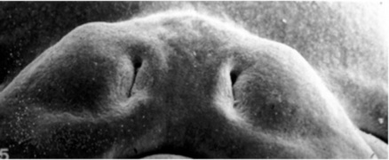
Embryonic Palate
|
Human primary palate |

|
- EM Links: Image - stage 16 | Image - stage 17 | Image - stage 18 | Image - stage 19
Fetal Palate
| Secondary palate, fusion in the human embryo in week 9. This requires the early palatal shelves growth, elevation, and fusion. There are many fusion events occurring during this period between each palatal shelf, to the primary palate, and also to the nasal septum.
palatal shelf elevation | secondary palate
|

Fetal Palate Growth (second trimester)[14] |

Fetal week 9 (GA week 11) hard palate fusion |

Ventral aspect of hard palate of human embryo of 80 mm |


Soft Palate
The soft palate mechanism of closure has not yet been determined, with several existing theories. A recent study[15] of embryos from the late embryonic-early fetal period (54 to 74 days post-conception) has identified the timing of soft palate closure.
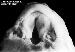
- 57 days - Late embryonic (Carnegie stage 23), epithelial seam present throughout the soft palate
- 64 days - Early fetal (Week 9), epithelium only persists in the most posterior regions of the soft palate
Postnatal Head Changes
Head Growth continues postnatally with fontanelle initially allowing head distortion on birth and early growth. These bony plates remain unfused to allow growth, puberty growth of face.
The palate also grows postnatally through childhood and becomes more elevated (arched) forming the "palatine vault", with different (but insignificant) growth between the genders.[16]
- Links: Postnatal Development
Animal Palate
Mouse Palate
- E11 - protrude from bilateral maxillary processes
- E12.5 - secondary palatal development begins
- E12.5-E14 - grow vertically along the developing tongue
- E14.5 - they elevate, meet, and fuse at the midline, to form an intact palate shelf, reflex opening and closing movements of the mouth
- E15.5 - palatal fusion is complete, mesenchymal condensation followed by osteogenic differentiation occurs.

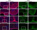
Mouse (E13.5) Palatal Shelf Wnt5a, Osr2 and Pax9 Expression.[17]
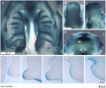
Image - Mouse E13.5 Bmp7 palate
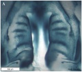
Image - palate
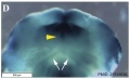
Image - palate detail


|

|
| Mouse ruga pattern (E16) | Mouse - Spry1 cleft palate |
- Links: Mouse Development | Bone Morphogenetic Protein | Wnt | Pax
Dog Palate

Newborn dog with cleft palate
Molecular
MMP25 PMID 20809987
Image - Mouse E13.5 Bmp7 palate PMID 23516636
Image - palate Bmp7 palate PMID 23516636
Image - palate detail Bmp7 palate PMID 23516636

- Links: Bone Morphogenetic Protein
Abnormalities
Note there are specific pages for both Cleft Lip and Palate and Cleft Palate.
Clinical Images
Cleft Lip/Palate
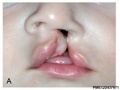
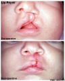
Unilateral cleft lip
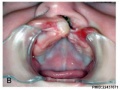
Unilateral cleft lip
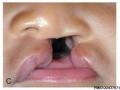
Complete Unilateral cleft lip and palate
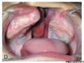
Complete Unilateral cleft lip and palate
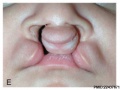
Complete bilateral cleft lip and palate
Complete bilateral cleft lip and palate
Cheiloplasty - surgical repair of the lip


Cleft Palate
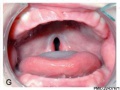
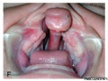
Incomplete cleft palate
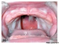
Cleft palate
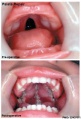
Palatoplasty - surgical repair of the palate


The way in which the upper jaw forms from fusion of the smaller upper prominence of the first pharyngeal arch leads to a common congenital defect in this region called "clefting", which may involve either the upper lip, the palate or both structures.
| Australian Palate Abnormalities (2002-2003) |
|---|
| Cleft lip with or without cleft palate (9.2 per 10,000 births) ICD-10 Q36.0, Q36.1, Q36.9, Q37.0–Q37.5, Q37.8, Q37.9 |
A congenital anomaly characterised by a partial or complete clefting of the upper lip, with or without clefting of the alveolar ridge or the hard palate. Excludes a midline cleft of the upper or lower lip and an oblique facial fissure (going towards the eye).
|
| Cleft palate without cleft lip (8.1 per 10,000 births) ICD-10 Q35.0–Q35.9 |
A congenital anomaly characterised by a closure defect of the hard and/or soft palate behind the foramen incisivum without a cleft lip. This anomaly includes sub-mucous cleft palate, but excludes cleft palate with a cleft lip, a functional short palate and high narrow palate.
|
|
| Global Orofacial Cleft Rate (1950 - 2015) | |||||||||||||
|---|---|---|---|---|---|---|---|---|---|---|---|---|---|
This data is from a study of the published data (1950 - 2015)[19]
|
International Classification of Diseases - Cleft Palate
Cleft lip and cleft palate (Q35-Q37)
Use additional code (Q30.2), if desired, to identify associated malformations of the nose. Excludes Robin's syndrome ( Q87.0 )
| Q37 | Cleft palate with cleft lip |
| Q37.0 | Cleft hard palate with bilateral cleft lip |
| Q37.1 | Cleft hard palate with unilateral cleft lip |
| Cleft hard palate with cleft lip NOS | |
| Q37.2 | Cleft soft palate with bilateral cleft lip |
| Q37.3 | Cleft soft palate with unilateral cleft lip |
| Cleft soft palate with cleft lip NOS | |
| Q37.4 | Cleft hard and soft palate with bilateral cleft lip |
| Q37.5 | Cleft hard and soft palate with unilateral cleft lip |
| Cleft hard and soft palate with cleft lip NOS | |
| Q37.8 | Unspecified cleft palate with bilateral cleft lip |
| Q37.9 | Unspecified cleft palate with unilateral cleft lip |
| Cleft palate with cleft lip NOS |
Embryonic Human Cleft Palate

|
| Stage16 (ventral view) |
Cleft Lip

|
|
| Cleft Lip Genes[20] | ||||||||||||||||||||||||||||||||||||||
|---|---|---|---|---|---|---|---|---|---|---|---|---|---|---|---|---|---|---|---|---|---|---|---|---|---|---|---|---|---|---|---|---|---|---|---|---|---|---|
Midline Cleft Lip Genes
| ||||||||||||||||||||||||||||||||||||||
|
Cleft Lip (+/− cleft palate) Genes
|
Cleft Palate


|
(Data: Congenital Malformations Australia 1981-1992 P. Lancaster and E. Pedisich ISSN 1321-8352)
Search Pubmed Now: cleft lip | cleft palate |
Cleft Risk Variants
Two genes were identified from a recent genome-wide study.[22]
- MAFB is expressed in the mouse palatal shelf.
- ABCA4 is a member of a superfamily of transmembrane proteins, and mutations in ABCA4 play a major role in the etiology of Stargardt disease and related retinopathies. Gene produces an ATP-binding cassette (ABC) superfamily trans-membrane protein
- Links: OMIM - MAFB | OMIM - ABCA4
Ten most frequently reported Birth Anomalies
- Hypospadias (More? Male movie | Genital Abnormalities - Hypospadia)
- Obstructive Defects of the Renal Pelvis (More? Renal System - Abnormalities)
- Ventricular Septal Defect (More? Cardiovascular Abnormalities - Ventricular Septal Defect)
- Congenital Dislocated Hip (More? Musculoskelal Abnormalities - Congenital Dislocation of the Hip (CDH))
- Trisomy 21 or Down syndrome - (More? Trisomy 21)
- Hydrocephalus (More? Hydrocephalus)
- Cleft Palate (More? Palate_Development)
- Trisomy 18 or Edward Syndrome - multiple abnormalities of the heart, diaphragm, lungs, kidneys, ureters and palate 86% discontinued (More? (More? Trisomy 18)
- Renal Agenesis/Dysgenesis - reduction in neonatal death and stillbirth since 1993 may be due to the more severe cases being identified in utero and being represented amongst the increased proportion of terminations (approximately 31%). (More? Renal System - Abnormalities)
- Cleft Lip and Palate - occur with another defect in 33.7% of cases.(More? Palate Development | Head Development)
(From the Victorian Perinatal Data Collection Unit in the Australian state of Victoria between 2003-2004)
Folate
A recent study of periconceptional folate supplementation using the Cochrane Pregnancy and Childbirth Group's Trials Register (July 2010) identified no statistically significant evidence of any effects on prevention of cleft palate and cleft lip at birth.[23]
References
- ↑ Xu J, Wang L, Li H, Yang T, Zhang Y, Hu T, Huang Z & Chen Y. (2019). Short stature homeobox 2 (SHOX2) regulates osteogenic differentiation and pattern formation during hard palate development in mice. J. Biol. Chem. , , . PMID: 31649032 DOI.
- ↑ Nakajima A, F Shuler C, Gulka AOD & Hanai JI. (2018). TGF-β Signaling and the Epithelial-Mesenchymal Transition during Palatal Fusion. Int J Mol Sci , 19, . PMID: 30463190 DOI.
- ↑ Dursun A, Öztürk K & Albay S. (2018). Development of Hard and Soft Palate During the Fetal Period and Hard Palate Asymmetry. J Craniofac Surg , 29, 2358-2362. PMID: 30320695 DOI.
- ↑ Hammond NL, Brookes KJ & Dixon MJ. (2018). Ectopic Hedgehog Signaling Causes Cleft Palate and Defective Osteogenesis. J. Dent. Res. , 97, 1485-1493. PMID: 29975848 DOI.
- ↑ Kishimoto H, Yamada S, Kanahashi T, Yoneyama A, Imai H, Matsuda T, Takeda T, Kawai K & Suzuki S. (2016). Three-dimensional imaging of palatal muscles in the human embryo and fetus: Development of levator veli palatini and clinical importance of the lesser palatine nerve. Dev. Dyn. , 245, 123-31. PMID: 26509917 DOI.
- ↑ Potter AS & Potter SS. (2015). Molecular Anatomy of Palate Development. PLoS ONE , 10, e0132662. PMID: 26168040 DOI.
- ↑ Mima J, Koshino A, Oka K, Uchida H, Hieda Y, Nohara K, Kogo M, Chai Y & Sakai T. (2013). Regulation of the epithelial adhesion molecule CEACAM1 is important for palate formation. PLoS ONE , 8, e61653. PMID: 23613893 DOI.
- ↑ Nelson ER, Levi B, Sorkin M, James AW, Liu KJ, Quarto N & Longaker MT. (2011). Role of GSK-3β in the osteogenic differentiation of palatal mesenchyme. PLoS ONE , 6, e25847. PMID: 22022457 DOI.
- ↑ San Miguel S, Serrano MJ, Sachar A, Henkemeyer M, Svoboda KK & Benson MD. (2011). Ephrin reverse signaling controls palate fusion via a PI3 kinase-dependent mechanism. Dev. Dyn. , 240, 357-64. PMID: 21246652 DOI.
- ↑ Beaty TH, Murray JC, Marazita ML, Munger RG, Ruczinski I, Hetmanski JB, Liang KY, Wu T, Murray T, Fallin MD, Redett RA, Raymond G, Schwender H, Jin SC, Cooper ME, Dunnwald M, Mansilla MA, Leslie E, Bullard S, Lidral AC, Moreno LM, Menezes R, Vieira AR, Petrin A, Wilcox AJ, Lie RT, Jabs EW, Wu-Chou YH, Chen PK, Wang H, Ye X, Huang S, Yeow V, Chong SS, Jee SH, Shi B, Christensen K, Melbye M, Doheny KF, Pugh EW, Ling H, Castilla EE, Czeizel AE, Ma L, Field LL, Brody L, Pangilinan F, Mills JL, Molloy AM, Kirke PN, Scott JM, Scott JM, Arcos-Burgos M & Scott AF. (2010). A genome-wide association study of cleft lip with and without cleft palate identifies risk variants near MAFB and ABCA4. Nat. Genet. , 42, 525-9. PMID: 20436469 DOI.
- ↑ Welsh IC, Hagge-Greenberg A & O'Brien TP. (2007). A dosage-dependent role for Spry2 in growth and patterning during palate development. Mech. Dev. , 124, 746-61. PMID: 17693063 DOI.
- ↑ Foppiano S, Hu D & Marcucio RS. (2007). Signaling by bone morphogenetic proteins directs formation of an ectodermal signaling center that regulates craniofacial development. Dev. Biol. , 312, 103-14. PMID: 18028903 DOI.
- ↑ Diewert VM & Lozanoff S. (1993). A morphometric analysis of human embryonic craniofacial growth in the median plane during primary palate formation. J. Craniofac. Genet. Dev. Biol. , 13, 147-61. PMID: 8227288
- ↑ BURDI AR. (1965). SAGITTAL GROWTH OF THE NASOMAXILLARY COMPLEX DURING THE SECOND TRIMESTER OF HUMAN PRENATAL DEVELOPMENT. J. Dent. Res. , 44, 112-25. PMID: 14245486 DOI.
- ↑ Danescu A, Mattson M, Dool C, Diewert VM & Richman JM. (2015). Analysis of human soft palate morphogenesis supports regional regulation of palatal fusion. J. Anat. , 227, 474-86. PMID: 26299693 DOI.
- ↑ Yang ST, Kim HK, Lim YS, Chang MS, Lee SP & Park YS. (2013). A three dimensional observation of palatal vault growth in children using mixed effect analysis: a 9 year longitudinal study. Eur J Orthod , 35, 832-40. PMID: 23314328 DOI.
- ↑ Almaidhan A, Cesario J, Landin Malt A, Zhao Y, Sharma N, Choi V & Jeong J. (2014). Neural crest-specific deletion of Ldb1 leads to cleft secondary palate with impaired palatal shelf elevation. BMC Dev. Biol. , 14, 3. PMID: 24433583 DOI.
- ↑ P. Lancaster and E. Pedisich, Congenital Malformations Australia 1981-1992, ISSN 1321-835.
- ↑ <pubmed>26742364</pubmed>
- ↑ 20.0 20.1 Dixon MJ, Marazita ML, Beaty TH & Murray JC. (2011). Cleft lip and palate: understanding genetic and environmental influences. Nat. Rev. Genet. , 12, 167-78. PMID: 21331089 DOI.
- ↑ Rastogi MV & LaFranchi SH. (2010). Congenital hypothyroidism. Orphanet J Rare Dis , 5, 17. PMID: 20537182 DOI.
- ↑ Cite error: Invalid
<ref>tag; no text was provided for refs namedPMID20436469 - ↑ De-Regil LM, Fernández-Gaxiola AC, Dowswell T & Peña-Rosas JP. (2010). Effects and safety of periconceptional folate supplementation for preventing birth defects. Cochrane Database Syst Rev , , CD007950. PMID: 20927767 DOI.
Journals
- The Cleft Palate-Craniofacial Journal Homepage | Available issues
Reviews
Indian J Plast Surg. 2009 October; 42(Suppl):Cleft Lip and Palate Issue
Nakajima A, F Shuler C, Gulka AOD & Hanai JI. (2018). TGF-β Signaling and the Epithelial-Mesenchymal Transition during Palatal Fusion. Int J Mol Sci , 19, . PMID: 30463190 DOI.
Tarr JT, Lambi AG, Bradley JP, Barbe MF & Popoff SN. (2018). Development of Normal and Cleft Palate: A Central Role for Connective Tissue Growth Factor (CTGF)/CCN2. J Dev Biol , 6, . PMID: 30029495 DOI.
Weng M, Chen Z, Xiao Q, Li R & Chen Z. (2018). A review of FGF signaling in palate development. Biomed. Pharmacother. , 103, 240-247. PMID: 29655165 DOI.
Lan Y, Xu J & Jiang R. (2015). Cellular and Molecular Mechanisms of Palatogenesis. Curr. Top. Dev. Biol. , 115, 59-84. PMID: 26589921 DOI.
Suzuki A, Sangani DR, Ansari A & Iwata J. (2016). Molecular mechanisms of midfacial developmental defects. Dev. Dyn. , 245, 276-93. PMID: 26562615 DOI.
Abramyan J & Richman JM. (2015). Recent insights into the morphological diversity in the amniote primary and secondary palates. Dev. Dyn. , 244, 1457-68. PMID: 26293818 DOI.
Bush JO & Jiang R. (2012). Palatogenesis: morphogenetic and molecular mechanisms of secondary palate development. Development , 139, 231-43. PMID: 22186724 DOI.
Meng L, Bian Z, Torensma R & Von den Hoff JW. (2009). Biological mechanisms in palatogenesis and cleft palate. J. Dent. Res. , 88, 22-33. PMID: 19131313 DOI.
Dudas M, Li WY, Kim J, Yang A & Kaartinen V. (2007). Palatal fusion - where do the midline cells go? A review on cleft palate, a major human birth defect. Acta Histochem. , 109, 1-14. PMID: 16962647 DOI.
Ferguson MW. (1988). Palate development. Development , 103 Suppl, 41-60. PMID: 3074914
Hay ED. (1995). An overview of epithelio-mesenchymal transformation. Acta Anat (Basel) , 154, 8-20. PMID: 8714286
Articles
Hammond NL, Brookes KJ & Dixon MJ. (2018). Ectopic Hedgehog Signaling Causes Cleft Palate and Defective Osteogenesis. J. Dent. Res. , 97, 1485-1493. PMID: 29975848 DOI.
Sun L, Wang J, Liu H, Fan Z, Wang S & Du J. (2017). A Comprehensive Study of Palate Development in Miniature Pig. Anat Rec (Hoboken) , 300, 1409-1419. PMID: 28296336 DOI.
Steding G & Jian Y. (2010). The origin and early development of the nasal septum in human embryos. Ann. Anat. , 192, 82-5. PMID: 20149609 DOI.
Xiong W, He F, Morikawa Y, Yu X, Zhang Z, Lan Y, Jiang R, Cserjesi P & Chen Y. (2009). Hand2 is required in the epithelium for palatogenesis in mice. Dev. Biol. , 330, 131-41. PMID: 19341725 DOI.
Search PubMed
Search Pubmed: palate development | cleft palate development |
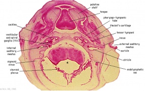
Additional Images
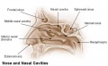
Nasal cavities and palate
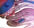
Palate, tongue and Meckel's cartilage
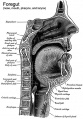
Historic Pharynx cartoon
Unilateral cleft lip and palate
Cleft palate feeder


Historic
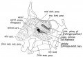
Fig. 6. Showing the structures formed in the Lateral Nasal Processes.
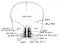
Fig. 7. Coronal section of the skull of a 7th month human foetus to show the cartilages of the Lateral and Mesial Nasal Processes and the bones formed round them.
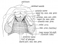
Fig. 8. Showing the ingrowth of the palatal plates of the two maxillary processes early in the 2nd month. (After Kollmann.) .
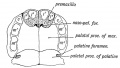
Fig. 9. Showing the Hard Palate at birth. The premaxillary part is formed from the Mesial Nasal Processes ; the remainder by the Palatal Plates of the Maxillary Processes.
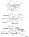
Fig. 10, a, b, c. Showing what become of the skeletons of the Mandibular Arch (Meckel's Cartilage) and Maxillary Process (Palato-quadrate Cartilage).
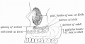
Fig. 11. Showing the manner in which the development of the Maxillary Antrum affects the size of the palate and position of the molar teeth.


Terms
| Palate Development (expand to see terms) |
|---|
|
| Other Terms Lists |
|---|
| Terms Lists: ART | Birth | Bone | Cardiovascular | Cell Division | Endocrine | Gastrointestinal | Genital | Genetic | Head | Hearing | Heart | Immune | Integumentary | Neonatal | Neural | Oocyte | Palate | Placenta | Radiation | Renal | Respiratory | Spermatozoa | Statistics | Tooth | Ultrasound | Vision | Historic | Drugs | Glossary |
External Links
External Links Notice - The dynamic nature of the internet may mean that some of these listed links may no longer function. If the link no longer works search the web with the link text or name. Links to any external commercial sites are provided for information purposes only and should never be considered an endorsement. UNSW Embryology is provided as an educational resource with no clinical information or commercial affiliation.
- Prof Virginia Diewert - Professor of Orthodontics, University of British Columbia, who recently visited the Lab and helped with content, organisation and development of the Palate Development section.
- NIH FACEBASE - Comprehensive craniofacial data and resources.
- Medline Plus - Cleft Lip and Palate
- Better Health Channel - Cleft palate and cleft lip
- March of Dimes Birth Defects Foundation - Cleft Palate
Glossary Links
- Glossary: A | B | C | D | E | F | G | H | I | J | K | L | M | N | O | P | Q | R | S | T | U | V | W | X | Y | Z | Numbers | Symbols | Term Link
Cite this page: Hill, M.A. (2024, June 10) Embryology Palate Development. Retrieved from https://embryology.med.unsw.edu.au/embryology/index.php/Palate_Development
- © Dr Mark Hill 2024, UNSW Embryology ISBN: 978 0 7334 2609 4 - UNSW CRICOS Provider Code No. 00098G