Category:Mouse E14.5

The Embryology pages and media listed below relate to mouse embryonic day 14.5 (E14.5) of development. This staging by "days" relate to in the female presence of a vaginal plug indicating that the mating occurred, see timed pregnancy.
- Mouse Stages: E1 | E2.5 | E3.0 | E3.5 | E4.5 | E5.0 | E5.5 | E6.0 | E7.0 | E7.5 | E8.0 | E8.5 | E9.0 | E9.5 | E10 | E10.5 | E11 | E11.5 | E12 | E12.5 | E13 | E13.5 | E14 | E14.5 | E15 | E15.5 | E16 | E16.5 | E17 | E17.5 | E18 | E18.5 | E19 | E20 | Timeline | About timed pregnancy
| Carnegie | Stage | |||||||||||||||||||||||
| Human | Days | 1 | 2-3 | 4-5 | 5-6 | 7-12 | 13-15 | 15-17 | 17-19 | 20 | 22 | 24 | 28 | 30 | 33 | 36 | 40 | 42 | 44 | 48 | 52 | 54 | 55 | 58 |
| Mouse | Days | 1 | 2 | 3 | E4.5 | E5.0 | E6.0 | E7.0 | E8.0 | E9.0 | E9.5 | E10 | E10.5 | E11 | E11.5 | E12 | E12.5 | E13 | E13.5 | E14 | E14.5 | E15 | E15.5 | E16 |
| Rat | Days | 1 | 3.5 | 4-5 | 5 | 6 | 7.5 | 8.5 | 9 | 10.5 | 11 | 11.5 | 12 | 12.5 | 13 | 13.5 | 14 | 14.5 | 15 | 15.5 | 16 | 16.5 | 17 | 17.5 |
| Note these Carnegie stages are only approximate day timings for average of embryos. Links: Carnegie Stage Comparison | ||||||||||||||||||||||||
| ||||||||||||||||||||||||
| Timeline Links: human timeline | mouse timeline | mouse detailed timeline | chicken timeline | rat timeline | Medaka | Category:Timeline |
Search Pubmed: Mouse E14.5
| Mouse Links: Introduction | Mouse Stages | Mouse Timeline | Mouse Timeline Detailed | Mouse Estrous Cycle | Mouse Heart | Mouse Knockout | Movie - Cephalic Plexus | Movie - Blastocyst Cdx2 | ANAT2341 Project 2009 | Category:Mouse | |||||||||||||||||||||||||||||||||||||||||||||||||||||||||||||||||||||||||||||||||||||||||||||||||||||||||||||||||||||||||||
|
| ||||||||||||||||||||||||||||||||||||||||||||||||||||||||||||||||||||||||||||||||||||||||||||||||||||||||||||||||||||||||||
Events
- Musculoskeletal System - Abdominal wall migration of myoblasts to the ventral midline almost complete, except panniculus carnosus. Directional organization of the myoblasts was observed in the external oblique, internal oblique, and transversus abdominis and myotubes appeared to be forming within these muscle groups. Connective tissue layers over the rectus abdominis are thicker than in earlier stages.[1]
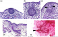
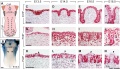
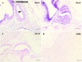
- palate - secondary palatal shelves elevate, meet, and begin to fuse at the midline, to form an intact palate shelf by Template:E15.5.
- reflex opening and closing movements of the mouth.
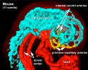
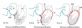
- Coronary Circulation - left and right coronary arteries developed.[2]
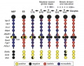
- thymus - [3] - Foxn1 (forkhead box N1) expression in the thymus domain of third pouch, hair follicles and the epidermis from about E14.5.
References
- ↑ Nichol PF, Corliss RF, Yamada S, Shiota K & Saijoh Y. (2012). Muscle patterning in mouse and human abdominal wall development and omphalocele specimens of humans. Anat Rec (Hoboken) , 295, 2129-40. PMID: 22976993 DOI.
- ↑ Red-Horse K, Ueno H, Weissman IL & Krasnow MA. (2010). Coronary arteries form by developmental reprogramming of venous cells. Nature , 464, 549-53. PMID: 20336138 DOI.
- ↑ Blackburn CC & Manley NR. (2004). Developing a new paradigm for thymus organogenesis. Nat. Rev. Immunol. , 4, 278-89. PMID: 15057786 DOI.
Search Pubmed: Mouse E14.5
Pages in category 'Mouse E14.5'
The following 3 pages are in this category, out of 3 total.
Media in category 'Mouse E14.5'
The following 38 files are in this category, out of 38 total.
 Anderson2016-fig27a.jpg 800 × 800; 117 KB
 Anderson2016-fig27b.jpg 800 × 800; 101 KB
 Anderson2016-fig33a.jpg 800 × 800; 110 KB
 Anderson2016-fig33b.jpg 800 × 800; 127 KB
 Anderson2016-fig46b.jpg 800 × 800; 120 KB
 Mouse - palate MMP-25 expression.jpg 1,000 × 818; 243 KB
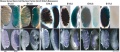
 Mouse bladder development E12.5-E16.5.jpg 1,105 × 1,000; 202 KB
 Mouse circumvallate papilla 01.jpg 712 × 955; 114 KB
 Mouse cochlea development cartoon.jpg 1,000 × 280; 53 KB
 Mouse cochlea gene expression.jpg 1,000 × 346; 75 KB
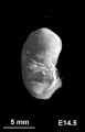 Mouse CT E14.5.jpg 221 × 344; 6 KB
 Mouse E14.5 gene expression.jpg 986 × 2,233; 259 KB
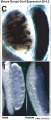
 Mouse E14.5 Titf1 gene expression.jpg 481 × 739; 33 KB
 Mouse embryo cortex radial expansion.jpg 1,200 × 624; 249 KB
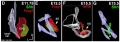
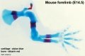
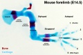
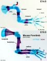 Mouse forelimb cartilage and bone E14.5 E18.5.jpg 1,000 × 1,300; 156 KB
 Mouse gonad Gcnf expression 01.jpg 1,947 × 843; 304 KB
 Mouse gonad Gcnf expression E14.5.jpg 334 × 784; 60 KB
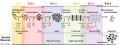 Mouse limb bone development timeline.jpg 1,256 × 469; 107 KB
 Mouse limb cartilage and bone E14.5.jpg 800 × 526; 42 KB
 Mouse limb cartilage and bone E14.5L.jpg 1,000 × 658; 73 KB
 Mouse limb tissue development.jpg 1,280 × 767; 161 KB
 Mouse lung development 01.jpg 1,000 × 1,254; 791 KB
 Mouse lung development 01a.jpg 800 × 1,003; 495 KB
 Mouse lung development 02.jpg 922 × 922; 239 KB
 Mouse lung development 03.jpg 540 × 1,200; 349 KB
 Mouse lung E14.5 Sox9.jpg 600 × 594; 48 KB
 Mouse mammary development 01.jpg 1,200 × 773; 147 KB
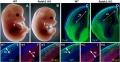
 Mouse melanoblast distribution 01.jpg 697 × 1,000; 192 KB
 Mouse melanoblast distribution 04.jpg 761 × 1,128; 177 KB
 Mouse pancreas development.jpg 600 × 939; 261 KB
 Mouse tongue Pax9 expression 01.jpg 1,200 × 687; 259 KB
 Mouse tongue Pax9 expression 02.jpg 1,200 × 833; 299 KB
 Mouse ventral body wall development 01.jpg 1,200 × 635; 130 KB
 Mouse whole lung E14.5.jpg 600 × 594; 17 KB
 Mouse- X-linked gene expression in primordial germ cells.jpg 800 × 632; 90 KB
 Mouse-coronary vessel formation.jpg 800 × 282; 44 KB
 Mouse-mammary-E14.5.jpg 600 × 459; 77 KB
 Mouse-pituitary Sox4 expression.jpg 596 × 448; 55 KB


