Vision - Cornea Development: Difference between revisions
(Created page with "{{Header}} ==Introduction== right|300px These notes introduce the development of the eye cornea. {{Vision Links}} {{Senses Links}} ==...") |
mNo edit summary |
||
| (37 intermediate revisions by the same user not shown) | |||
| Line 1: | Line 1: | ||
{{Header}} | {{Header}} | ||
==Introduction== | ==Introduction== | ||
[[File: | [[File:Mouse eye neural crest cornea 01.jpg||thumb|300px|alt=Cornea structure]] | ||
These notes introduce the development of the eye cornea. | [[File:Stage_22_image_208.jpg|thumb|300px|alt=Human embryonic cornea|Human embryonic cornea ([[Week 8]], [[Carnegie stage 22]])]] | ||
[[File:Stage_22_image_212.jpg|thumb|300px|alt=Human embryonic cornea|Human embryonic cornea detail ([[Week 8]], [[Carnegie stage 22]])]] | |||
[[File:Gray0883.jpg|thumb|300px|alt=Section through front of Eyeball|Section through human cornea]] | |||
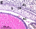
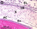
These notes introduce the development of the cornea of the eye. The adult cornea has three layers: an outer epithelium layer (ectoderm), a middle stromal layer of collagen-rich extracellular matrix between stromal keratocytes (neural crest) and an inner layer of endothelial cells (neural crest). | |||
The cornea is a vision-specific specialised sensory epithelia that in humans differentiates mainly in the postnatal period. It arises initially from cranial ectoderm adjacent to the lens placode and forms a presumptive corneal epithelium. Later neural crest cells migrate between the lens and presumptive structure to form both the corneal endothelium and the stromal fibroblasts (keratocytes). Neural crest development in humans, reptiles and birds differs from that seen in rodents, cats, rabbits, and cattle. | |||
{{ | {{Vision Links}} | ||
:'''Links:''' [[:Category:Cornea|Category:Cornea]] | [[Neural Crest Development]] | [[Integumentary Development]] | |||
== Some Recent Findings == | == Some Recent Findings == | ||
{| | {| | ||
|-bgcolor="F5FAFF" | |-bgcolor="F5FAFF" | ||
| | | | ||
* ''' | * '''Review - Corneal Development Different Cells from a Common Progenitor'''{{#pmid:26310148|PMID26310148}} "Development of the vertebrate cornea is a multistep process that involves cellular interactions between various ectodermal-derived tissues. Bilateral interactions between the neural ectoderm-derived optic vesicles and the cranial ectoderm give rise to the presumptive corneal epithelium and other epithelia of the ocular surface. Interactions between the neural tube and the adjacent ectoderm give rise to the neural crest cells, a highly migratory and multipotent cell population. Neural crest cells migrate between the lens and presumptive corneal epithelium to form the corneal endothelium and the stromal keratocytes. The sensory nerves that abundantly innervate the corneal stroma and epithelium originate from the neural crest- and ectodermal placode-derived trigeminal ganglion." | ||
* ''' | |||
* '''Bovine cornea extracellular matrix structure'''{{#pmid:25019467|PMID25019467}} "Electron microscopy and X-ray fibre diffraction were used to ascertain collagen fibril architecture. The bovine cornea was 1021±5.42μm thick at its outer periphery, defined as 9-12mm from the corneal centre, compared to 844±8.10μm at the centre. The outer periphery of the cornea was marginally, but not significantly, more hydrated than the centre (H=4.3 vs. H=3.7), and was more abundant in hydroxyproline (0.12 vs. 0.06mg/mg dry weight of cornea). DMMB assays indicated no change in the total amount of sulphated GAG across the cornea. Immunohistochemistry revealed the presence of both high- and low-sulphated epitopes of KS, as well as DS, throughout the cornea, and CS only in the peripheral cornea before the limbus. Quantification by ELISA, disclosed that although both high- and low-sulphated KS remained constant throughout stromal depth at different radial positions, high-sulphated epitopes remained constant from the corneal centre to outer-periphery, whereas low-sulphated epitopes increased significantly. | |||
|} | |} | ||
{| class="wikitable mw-collapsible mw-collapsed" | {| class="wikitable mw-collapsible mw-collapsed" | ||
! More recent papers | ! More recent papers | ||
|- | |- | ||
| [[File:Mark_Hill.jpg|90px|left]] {{Most_Recent_Refs}} | | [[File:Mark_Hill.jpg|90px|left]] {{Most_Recent_Refs}} | ||
Search term: '' | Search term: [http://www.ncbi.nlm.nih.gov/pubmed/?term=Cornea+Development ''Cornea Development''] | ||
<pubmed limit=5>Cornea Development</pubmed> | |||
Search term: [http://www.ncbi.nlm.nih.gov/pubmed/?term=Cornea+Embryology ''Cornea Embryology''] | |||
<pubmed limit=5> | <pubmed limit=5>Cornea Embryology</pubmed> | ||
|} | |} | ||
==Timeline== | ==Carnegie Stages - Eye== | ||
{{Eye Timeline table}} | |||
==Human Cornea== | |||
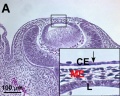
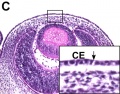
===Week 8 Stage 22=== | |||
The images below link to virtual slides of the human developing eye at [[Carnegie stage 22]]. Click on the image to open or select specific regions from the regions of interest links. | |||
{| | {| | ||
| | | {{SlideStage22-08}} | ||
| {{SlideStage22-08-eye}} | |||
* | | | ||
* | ====Virtual Slide - Regions of Interest==== | ||
* [http://embryology.med.unsw.edu.au/embryology/Slides/Embryo_Stages/Stage22/08-eye/Stage22-08-eye.html?zoom=3&lat=-3544&lon=4688&layers=B Eye Overview] | |||
* | * [http://embryology.med.unsw.edu.au/embryology/Slides/Embryo_Stages/Stage22/08-eye/Stage22-08-eye.html?zoom=4&lat=-2030&lon=2572&layers=B Lens and Cornea] | ||
* | * [http://embryology.med.unsw.edu.au/embryology/Slides/Embryo_Stages/Stage22/08-eye/Stage22-08-eye.html?zoom=5&lat=-1208&lon=2473&layers=B Cornea and Anterior Chamber] | ||
| [[File: | * [http://embryology.med.unsw.edu.au/embryology/Slides/Embryo_Stages/Stage22/08-eye/Stage22-08-eye.html?zoom=6&lat=-2410&lon=1843&layers=B Iris and Lens] | ||
|- | |||
|} | |||
'''Links:''' [[Embryo Virtual Slides]] | |||
==Cornea Epithelia== | |||
{| | |||
| The cornea ocular surface is composed of three epithelia, conjunctival, limbal and corneal. | |||
* Limbal stem cells are located in the palisades of Vogt, the transitional zone between the cornea and the conjunctiva. | |||
* Limbal stem cells are close to blood vessels. | |||
* They generate transient amplifying cells that terminally differentiate after a discrete number of cell divisions to corneal epithelial cells and undergo both centripetal migration and vertical migration. | |||
| [[File:Corneal_epithelial_cells_01.jpg|alt=Corneal Epithelial Cells cartoon|300px]] | |||
Corneal epithelial cells cartoon{{#pmid:19668514|PMID19668514}} | |||
|- | |||
| The Adult Human Limbal Palisades of Vogt | |||
* '''A''' - Palisades of Vogt (arrow) are readily recognized in the human limbus. | |||
* '''B''' - Such a unique pigmented structure can be identified on the flat mount preparation of Dispase-isolated human limbal epithelial sheets. | |||
* '''C''' - In donors with a darker skin, these palisades of Vogt are pigmented (arrow). | |||
* '''D''' - Under higher magnification, these limbal areas show undulated epithelial papillae (stars). | |||
* '''E''' - Hematoxyline staining highlights higher stratification and more undulation of the limbal epithelium, and the underlying limbal stroma has high cellularity and vascularity (arrow shows blood vessel). | |||
Bar represents 500 μm in A and B, 200 μm in C and E, and 50 μm in D | |||
| [[File:Limbal palisades of Vogt PMID17211449.jpg|alt=Adult human limbal palisades of Vogt|300px]] | |||
Adult human limbal palisades of Vogt{{#pmid:17211449|PMID17211449}} | |||
|} | |} | ||
=== | ===Limbal Stem Cells=== | ||
[[File:Limbal_stem_cell_niche_cartoon_PMID17211449.jpg|500px]] | |||
Cartoon showing the location of limbal stem cells at the limbal basal layer.{{#pmid:17211449|PMID17211449}} | |||
:'''Links:''' [[Stem Cells]] | |||
[[ | |||
==Descemet Membrane== | |||
Corneal endothelium basement membrane beginning in children at 3 μm thick and increases in adults to 10 μm. Consists of collagen type IV and VIII fibrils. | |||
: | Composed of two layers: | ||
# anterior banded layer - commencing in week 10 ({{GA}} week 12) as collagen lamellae and proteoglycans. | |||
# posterior non-banded layer - deposited by endothelial cells over time and thickens postnatally over decades. | |||
Descemet membrane was historically named after Jean Descemet (1732–1810) a French physician. | |||
==Palisades of Vogt== | |||
The palisades of Vogt are a series of radially oriented fibrovascular ridges concentrated along the upper and lower corneoscleral limbus, the vasculature component consists of radially oriented hairpin loops of narrow arterial and venous vessels. Named by Vogt in 1921. (for review see{{#pmid:7182957|PMID7182957}}) | |||
Aggregate into distinct crescentic zones and lie peripheral to the terminal capillary loops of the limbus and central to Schlemm’s canal. Lying between the connective tissue palisades are intervening radial zones of thickened conjunctival epithelium, the so-called inter-palisades or epithelial rete ridges. | |||
==Mouse Cornea== | |||
{| | {| | ||
| [[File:Mouse eye neural crest.jpg|400px]] | | [[File:Mouse eye neural crest.jpg|alt=histology Mouse eye neural crest|400px]] | ||
Neural crest-derived cells contribute to mouse cornea development.{{#pmid:16403239|PMID16403239}} | |||
| | |||
* '''a''' Toluidine blue staining of an adult eye. The boxed areas correspond to b and c | |||
* '''b''' A detailed view of the corneal assembly, including outer epithelium, stroma, and inner endothelium | |||
* '''c''' The chamber angle at the irido-corneal transition which includes the trabecular meshwork (tm). | |||
* '''d-j''' In vivo fate mapping of NC-derived, β-galactosidase (βGal)-expressing cells (blue) | |||
* '''d''' The NC origin of corneal keratocytes (arrows) and of corneal endothelium (arrowhead). | |||
* '''e''' Structures of the chamber angle, including the trabecular meshwork are seen to be NC-derived. | |||
* '''f''' At E10, the optic cup is surrounded by NC-derived cells expressing βGal. | |||
* '''g-i''' The majority of the cells in the periocular mesenchyme (arrows), which forms the anterior eye segment, are of NC origin, as assessed from E11.5 to E13.5. | |||
* '''j''' The primary vitreous at E13.5 (arrowheads) shows a strong NC contribution. | |||
|} | |||
<gallery> | |||
File:Mouse cornea E12.5.jpg|Mouse E12.5 | |||
File:Mouse cornea E13.5.jpg|Mouse E13.5 | |||
File:Mouse cornea E16.5.jpg|Mouse E16.5 | |||
File:Mouse cornea P0.jpg|Mouse P0 | |||
</gallery> | |||
==Frog Cornea== | |||
This developmental timeline is from a recent frog (Xenopus laevis) cornea study{{#pmid:23896054|PMID23896054}} | |||
* '''stage 25''' - cornea starts from a simple embryonic epidermis overlying the developing optic vesicle. | |||
* '''stage 30''' - detachment of the lens placode, cranial neural crest cells start to invade the space between the lens and the embryonic epidermis to construct the corneal endothelium. | |||
* '''stage 41''' - a second wave of migratory cells containing presumptive keratocytes invades the matrix leading to the formation of inner cornea and outer cornea. A unique cell mass (stroma attracting center) connects the two layers like the center pole of a tent. | |||
* '''stage 48''' - many secondary stromal keratocytes individually migrate to the center and form the stroma layer. | |||
* '''stage 60''' - the stroma space is filled by collagen lamellae and keratocytes, and the stroma attracting center disappears. At early metamorphosis, the embryonic epithelium gradually changes to the adult corneal epithelium, which is covered by microvilli. | |||
* '''stage 62''' - the embryonic epithelium thickens and cell death is observed in the epithelium, coinciding with eyelid opening. | |||
* '''After metamorphosis''' - cornea has attained the adult structure of three cellular layers, epithelium, stroma, and endothelium, and between the cellular layers lie two acellular layers (Bowman's layer and Descemet's membrane) | |||
[[File:Xenopus_cornea_development_timeline.jpg|800px||alt=Xenopus cornea development timeline]] | |||
:'''Links:''' [[ | :'''Links:''' [[Frog Development]] | ||
==Molecular== | |||
[[File:Mouse eye TGF-beta model.jpg|600px]] | |||
Mouse Eye TGF-beta Model - Summary of the TGFβ-dependent development of anterior and posterior ocular structures.{{#pmid:16403239|PMID16403239}} | |||
{| | {| | ||
| valign=top|'''a''' Neural crest-derived cells (NC, blue) contribute to structures of the anterior eye segment and the primary vitreous (PV). | |||
* TGFβ signaling is involved in the formation of the ciliary body (CB) and the trabecular meshwork (TM), and in control of PV growth. | |||
* Moreover, normal PV development and/or TGFβ signaling are important for correct retinal patterning. | |||
| | | valign=top|'''b''' In the cornea, prospective stromal keratocytes and endothelial cells are of neural crest origin. | ||
* Here, TGFβ signaling is needed for the expression of the transcription factors Foxc1 and Pitx2 and for normal differentiation of NC-derived cells into collagen-synthesizing stromal keratocytes. | |||
* | * Moreover, in forming corneal endothelial cells (and in the TM), expression of Foxc1 and cell survival requires TGFβ signalling. | ||
* | |||
| valign=top | |||
* | |||
* | |||
|} | |} | ||
==Additional Images== | ==Additional Images== | ||
<gallery> | <gallery> | ||
</gallery> | </gallery> | ||
===Historic Images=== | ===Historic Images=== | ||
<gallery> | <gallery> | ||
File: | File:Foster052.jpg|Fig. 52. Eye of a Fowl on the day 8 | ||
File:Foster128.jpg|Fig. 128. Eye of a Rabbit Embryo 12 Days | |||
File: | File:Kollmann713.jpg| | ||
File: | File:Kollmann715.jpg| | ||
File: | File:Kollmann717.jpg| | ||
File: | |||
File:Bailey463.jpg|Fig. 463. Developing lens and optic cup. | File:Bailey463.jpg|Fig. 463. Developing lens and optic cup. | ||
File:Bailey464.jpg|Fig. 464. Model showing lens and formation of optic cup. | File:Bailey464.jpg|Fig. 464. Model showing lens and formation of optic cup. | ||
| Line 141: | Line 184: | ||
File:Bailey466.jpg|Fig. 466. Section through optic cup and lens invagination of chick of fifty-four hours' incubation. | File:Bailey466.jpg|Fig. 466. Section through optic cup and lens invagination of chick of fifty-four hours' incubation. | ||
File:Bailey467.jpg|Fig. 467. Section through eye of human embryo of 13-14 weeks. | File:Bailey467.jpg|Fig. 467. Section through eye of human embryo of 13-14 weeks. | ||
File:Brown001.jpg|Fig. 1. Section through head of pig, 2 mm long. | File:Brown001.jpg|Fig. 1. Section through head of pig, 2 mm long. | ||
File:Brown002.jpg|Fig. 2. Section through head of chick, 2 mm long. | File:Brown002.jpg|Fig. 2. Section through head of chick, 2 mm long. | ||
| Line 154: | Line 194: | ||
File:Brown009.jpg|Fig. 9. Section through head of pig, 9 mm long. | File:Brown009.jpg|Fig. 9. Section through head of pig, 9 mm long. | ||
File:Brown010.jpg | File:Brown010.jpg | ||
</gallery> | </gallery> | ||
| Line 160: | Line 199: | ||
<references/> | <references/> | ||
=== | ===Journals=== | ||
* | * [http://journals.lww.com/corneajrnl/pages/default.aspx Cornea] "For corneal specialists and for all general ophthalmologists with an interest in this exciting subspecialty, Cornea brings together the latest clinical and basic research on the cornea and the anterior segment of the eye." [http://www.ncbi.nlm.nih.gov/pubmed?term=%22Cornea%22[jour] PuMed Listing] | ||
===Reviews=== | |||
{{#pmid:26310148}} | |||
{{#pmid:23819758}} | |||
{{#pmid:20599432}} | |||
{{#pmid:19343693}} | |||
The International Journal of Developmental Biology [http://www.ijdb.ehu.es/web/contents.php?vol=48&issue=8-9 Vol. 48 Nos. 8/9 (2004) Eye Development] | The International Journal of Developmental Biology [http://www.ijdb.ehu.es/web/contents.php?vol=48&issue=8-9 Vol. 48 Nos. 8/9 (2004) Eye Development] | ||
===Articles=== | ===Articles=== | ||
{{#pmid:13429485}} | |||
'''Bookshelf''' [http://www.ncbi.nlm.nih.gov/sites/entrez?db=Books&cmd=search&term=cornea%20development cornea development] | |||
'''Bookshelf''' [http://www.ncbi.nlm.nih.gov/sites/entrez?db=Books&cmd=search&term= | |||
===Search Pubmed=== | ===Search Pubmed=== | ||
'''Search Pubmed:''' [http://www.ncbi.nlm.nih.gov/pubmed?term= | '''Search Pubmed:''' [http://www.ncbi.nlm.nih.gov/pubmed?term=cornea%20development cornea development] | ||
'''Search Entrez:''' [http://www.ncbi.nlm.nih.gov/sites/gquery?itool=toolbar&cmd=search&term= | '''Search Entrez:''' [http://www.ncbi.nlm.nih.gov/sites/gquery?itool=toolbar&cmd=search&term=cornea%20development cornea development] | ||
==Terms== | ==Terms== | ||
* '''Limbal epithelial stem cells''' - cells located at the limbal basal layer. | |||
* '''palisades of Vogt''' - series of radially oriented fibrovascular ridges concentrated along the upper and lower corneoscleral limbus, the vasculature component consists of radially oriented hairpin loops of narrow arterial and venous vessels. Named by Vogt in 1921. [https://www.ncbi.nlm.nih.gov/pubmed/7182957 PMID 7182957] | |||
. | |||
==External Links== | ==External Links== | ||
{{External Links}} | {{External Links}} | ||
* UNSW SoMS research - [https://medicalsciences.med.unsw.edu.au/research/groups/mechanisms-disease-and-translational-research MDTR] | |||
* '''UNSW Virtual Slides''' [http://vslides.unsw.edu.au/VirtualSlideV2.nsf/id/D0C49C Eye Development Histology] (requires login) | * '''UNSW Virtual Slides''' [http://vslides.unsw.edu.au/VirtualSlideV2.nsf/id/D0C49C Eye Development Histology] (requires login) | ||
{{Glossary}} | {{Glossary}} | ||
{{Footer}} | {{Footer}} | ||
[[Category:Vision]] | [[Category:Vision]][[Category:Cornea]][[Category:Neural Crest]] | ||
Latest revision as of 10:34, 9 August 2018
| Embryology - 25 May 2024 |
|---|
| Google Translate - select your language from the list shown below (this will open a new external page) |
|
العربية | català | 中文 | 中國傳統的 | français | Deutsche | עִברִית | हिंदी | bahasa Indonesia | italiano | 日本語 | 한국어 | မြန်မာ | Pilipino | Polskie | português | ਪੰਜਾਬੀ ਦੇ | Română | русский | Español | Swahili | Svensk | ไทย | Türkçe | اردو | ייִדיש | Tiếng Việt These external translations are automated and may not be accurate. (More? About Translations) |
Introduction


These notes introduce the development of the cornea of the eye. The adult cornea has three layers: an outer epithelium layer (ectoderm), a middle stromal layer of collagen-rich extracellular matrix between stromal keratocytes (neural crest) and an inner layer of endothelial cells (neural crest).
The cornea is a vision-specific specialised sensory epithelia that in humans differentiates mainly in the postnatal period. It arises initially from cranial ectoderm adjacent to the lens placode and forms a presumptive corneal epithelium. Later neural crest cells migrate between the lens and presumptive structure to form both the corneal endothelium and the stromal fibroblasts (keratocytes). Neural crest development in humans, reptiles and birds differs from that seen in rodents, cats, rabbits, and cattle.
Some Recent Findings
|
| More recent papers |
|---|
 This table allows an automated computer search of the external PubMed database using the listed "Search term" text link.
More? References | Discussion Page | Journal Searches | 2019 References | 2020 References Search term: Cornea Development <pubmed limit=5>Cornea Development</pubmed> Search term: Cornea Embryology <pubmed limit=5>Cornea Embryology</pubmed> |
Carnegie Stages - Eye
| Carnegie Stage | Event | ||||||||||||||||||
|---|---|---|---|---|---|---|---|---|---|---|---|---|---|---|---|---|---|---|---|
| 10 | optic primordia appear. | ||||||||||||||||||
| 11 | Right and left optic primordia meet at the optic chiasma forming a U-shaped rim. | ||||||||||||||||||
| 12 | optic neural crest reaches its maximum extent and the optic vesicle becomes covered by a complete sheath, | ||||||||||||||||||
| 13 | By the end of the fourth week the optic vesicle lies close to the surface ectoderm. Optic evagination differentiation allows identification of optic part of retina, future pigmented layer of retina, and optic stalk. The surface ectoderm overlying the optic vesicle, in response to this contact, has thickened to form the lense placode. | ||||||||||||||||||
| 14 | (about 32 days) Lens placode is indented by the lens pit, cup-shaped and still communicates with the surface by a narrowing pore. | ||||||||||||||||||
| 15 | (about 33 days) Lens pit is closed. The lens vesicle and optic cup lie close to the surface ectoderm and appear to press against the surface. | ||||||||||||||||||
| 16 | (37 days) Growth of the lens body results in a D-shaped lens cavity. Perilental blood vessels (tunica vasculosa lentis) are visible. Prior to the development of the eyelids, one small sulcus or groove forms above the eye (eyelid groove) and another below it. | ||||||||||||||||||
| 17 - 19 | Retinal pigment is visible and the retinal fissure is largely closed. Eyelids grooves deepen, eyelid folds develop, first below, and then above, the eye. | ||||||||||||||||||
| 18 | Mesenchyme invades the region between the lens epithelium and the surface ectoderm. | ||||||||||||||||||
| 19 - 22 | Eyelid folds develop into the eyelids and cover more of the eye as the palpebral fissure takes shape. The upper and the lower eyelids meet at the outer canthus in Stage 19. | ||||||||||||||||||
| 20 | Lens cavity is lost and a lens suture begins to form. The inner canthus is established. | ||||||||||||||||||
| 23 | retina comprises the pigmented layer, external limiting membrane, proliferative zone, external neuroblastic layer, transient fiber layer, internal neuroblastic layer, nerve fiber layer, and internal limiting membrane. Eyelids closure is complete (Note - shown as still open in the Kyoto embryo). | ||||||||||||||||||
Data from a study of human embryonic carnegie stages[3] and other sources.
| |||||||||||||||||||
Human Cornea
Week 8 Stage 22
The images below link to virtual slides of the human developing eye at Carnegie stage 22. Click on the image to open or select specific regions from the regions of interest links.
|
|
Virtual Slide - Regions of Interest |


Links: Embryo Virtual Slides
Cornea Epithelia
The cornea ocular surface is composed of three epithelia, conjunctival, limbal and corneal.
|

Corneal epithelial cells cartoon[4] |
The Adult Human Limbal Palisades of Vogt
Bar represents 500 μm in A and B, 200 μm in C and E, and 50 μm in D
|

Adult human limbal palisades of Vogt[5] |
Limbal Stem Cells

Cartoon showing the location of limbal stem cells at the limbal basal layer.[5]
- Links: Stem Cells
Descemet Membrane
Corneal endothelium basement membrane beginning in children at 3 μm thick and increases in adults to 10 μm. Consists of collagen type IV and VIII fibrils.
Composed of two layers:
- anterior banded layer - commencing in week 10 (GA week 12) as collagen lamellae and proteoglycans.
- posterior non-banded layer - deposited by endothelial cells over time and thickens postnatally over decades.
Descemet membrane was historically named after Jean Descemet (1732–1810) a French physician.
Palisades of Vogt
The palisades of Vogt are a series of radially oriented fibrovascular ridges concentrated along the upper and lower corneoscleral limbus, the vasculature component consists of radially oriented hairpin loops of narrow arterial and venous vessels. Named by Vogt in 1921. (for review see[6])
Aggregate into distinct crescentic zones and lie peripheral to the terminal capillary loops of the limbus and central to Schlemm’s canal. Lying between the connective tissue palisades are intervening radial zones of thickened conjunctival epithelium, the so-called inter-palisades or epithelial rete ridges.
Mouse Cornea

Neural crest-derived cells contribute to mouse cornea development.[7] |
|
Mouse E12.5
Mouse E13.5
Mouse E16.5
Mouse P0


Frog Cornea
This developmental timeline is from a recent frog (Xenopus laevis) cornea study[8]
- stage 25 - cornea starts from a simple embryonic epidermis overlying the developing optic vesicle.
- stage 30 - detachment of the lens placode, cranial neural crest cells start to invade the space between the lens and the embryonic epidermis to construct the corneal endothelium.
- stage 41 - a second wave of migratory cells containing presumptive keratocytes invades the matrix leading to the formation of inner cornea and outer cornea. A unique cell mass (stroma attracting center) connects the two layers like the center pole of a tent.
- stage 48 - many secondary stromal keratocytes individually migrate to the center and form the stroma layer.
- stage 60 - the stroma space is filled by collagen lamellae and keratocytes, and the stroma attracting center disappears. At early metamorphosis, the embryonic epithelium gradually changes to the adult corneal epithelium, which is covered by microvilli.
- stage 62 - the embryonic epithelium thickens and cell death is observed in the epithelium, coinciding with eyelid opening.
- After metamorphosis - cornea has attained the adult structure of three cellular layers, epithelium, stroma, and endothelium, and between the cellular layers lie two acellular layers (Bowman's layer and Descemet's membrane)

- Links: Frog Development
Molecular

Mouse Eye TGF-beta Model - Summary of the TGFβ-dependent development of anterior and posterior ocular structures.[7]
a Neural crest-derived cells (NC, blue) contribute to structures of the anterior eye segment and the primary vitreous (PV).
|
b In the cornea, prospective stromal keratocytes and endothelial cells are of neural crest origin.
|
Additional Images
Historic Images
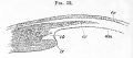
Fig. 52. Eye of a Fowl on the day 8
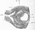
Fig. 128. Eye of a Rabbit Embryo 12 Days
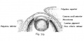
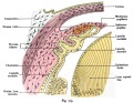
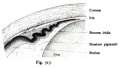
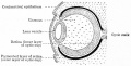
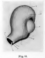
Fig. 463. Developing lens and optic cup.
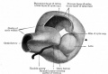
Fig. 464. Model showing lens and formation of optic cup.

Fig. 465. Stages in the development of the lens in the rabbit embryo.
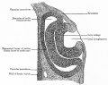
Fig. 466. Section through optic cup and lens invagination of chick of fifty-four hours' incubation.
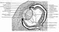
Fig. 467. Section through eye of human embryo of 13-14 weeks.
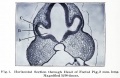
Fig. 1. Section through head of pig, 2 mm long.
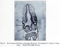
Fig. 2. Section through head of chick, 2 mm long.
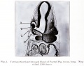
Fig. 3. Section through head of Foetal Pig, 2 mm long.
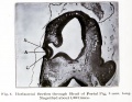
Fig. 4. Section through head of Foetal Pig, 3 mm long.
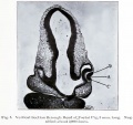
Fig. 5. Section through head of Foetal Pig, 3 mm long.
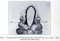
Fig. 6. Section through head of Foetal Pig, 4 mm long.
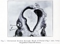
Fig. 7. Section through head of Foetal Pig, 7 mm long.
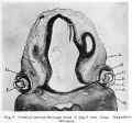
Fig. 8. Section through head of pig, 8 mm long.
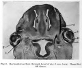
Fig. 9. Section through head of pig, 9 mm long.


References
- ↑ Lwigale PY. (2015). Corneal Development: Different Cells from a Common Progenitor. Prog Mol Biol Transl Sci , 134, 43-59. PMID: 26310148 DOI.
- ↑ Ho LT, Harris AM, Tanioka H, Yagi N, Kinoshita S, Caterson B, Quantock AJ, Young RD & Meek KM. (2014). A comparison of glycosaminoglycan distributions, keratan sulphate sulphation patterns and collagen fibril architecture from central to peripheral regions of the bovine cornea. Matrix Biol. , 38, 59-68. PMID: 25019467 DOI.
- ↑ Pearson AA. (1980). The development of the eyelids. Part I. External features. J. Anat. , 130, 33-42. PMID: 7364662
- ↑ Kayama M, Kurokawa MS, Ueno H & Suzuki N. (2007). Recent advances in corneal regeneration and possible application of embryonic stem cell-derived corneal epithelial cells. Clin Ophthalmol , 1, 373-82. PMID: 19668514
- ↑ 5.0 5.1 Li W, Hayashida Y, Chen YT & Tseng SC. (2007). Niche regulation of corneal epithelial stem cells at the limbus. Cell Res. , 17, 26-36. PMID: 17211449 DOI.
- ↑ Goldberg MF & Bron AJ. (1982). Limbal palisades of Vogt. Trans Am Ophthalmol Soc , 80, 155-71. PMID: 7182957
- ↑ 7.0 7.1 Ittner LM, Wurdak H, Schwerdtfeger K, Kunz T, Ille F, Leveen P, Hjalt TA, Suter U, Karlsson S, Hafezi F, Born W & Sommer L. (2005). Compound developmental eye disorders following inactivation of TGFbeta signaling in neural-crest stem cells. J. Biol. , 4, 11. PMID: 16403239 DOI.
- ↑ Hu W, Haamedi N, Lee J, Kinoshita T & Ohnuma S. (2013). The structure and development of Xenopus laevis cornea. Exp. Eye Res. , 116, 109-28. PMID: 23896054 DOI.
Journals
- Cornea "For corneal specialists and for all general ophthalmologists with an interest in this exciting subspecialty, Cornea brings together the latest clinical and basic research on the cornea and the anterior segment of the eye." [jour PuMed Listing]
Reviews
Lwigale PY. (2015). Corneal Development: Different Cells from a Common Progenitor. Prog Mol Biol Transl Sci , 134, 43-59. PMID: 26310148 DOI.
Maycock NJ & Marshall J. (2014). Genomics of corneal wound healing: a review of the literature. Acta Ophthalmol , 92, e170-84. PMID: 23819758 DOI.
Hassell JR & Birk DE. (2010). The molecular basis of corneal transparency. Exp. Eye Res. , 91, 326-35. PMID: 20599432 DOI.
Masters BR. (2009). Correlation of histology and linear and nonlinear microscopy of the living human cornea. J Biophotonics , 2, 127-39. PMID: 19343693 DOI.
The International Journal of Developmental Biology Vol. 48 Nos. 8/9 (2004) Eye Development
Articles
MAURICE DM. (1957). The structure and transparency of the cornea. J. Physiol. (Lond.) , 136, 263-86. PMID: 13429485
Bookshelf cornea development
Search Pubmed
Search Pubmed: cornea development
Search Entrez: cornea development
Terms
- Limbal epithelial stem cells - cells located at the limbal basal layer.
- palisades of Vogt - series of radially oriented fibrovascular ridges concentrated along the upper and lower corneoscleral limbus, the vasculature component consists of radially oriented hairpin loops of narrow arterial and venous vessels. Named by Vogt in 1921. PMID 7182957
.
External Links
External Links Notice - The dynamic nature of the internet may mean that some of these listed links may no longer function. If the link no longer works search the web with the link text or name. Links to any external commercial sites are provided for information purposes only and should never be considered an endorsement. UNSW Embryology is provided as an educational resource with no clinical information or commercial affiliation.
- UNSW SoMS research - MDTR
- UNSW Virtual Slides Eye Development Histology (requires login)
Glossary Links
- Glossary: A | B | C | D | E | F | G | H | I | J | K | L | M | N | O | P | Q | R | S | T | U | V | W | X | Y | Z | Numbers | Symbols | Term Link
Cite this page: Hill, M.A. (2024, May 25) Embryology Vision - Cornea Development. Retrieved from https://embryology.med.unsw.edu.au/embryology/index.php/Vision_-_Cornea_Development
- © Dr Mark Hill 2024, UNSW Embryology ISBN: 978 0 7334 2609 4 - UNSW CRICOS Provider Code No. 00098G