File:Pituitary histology 001.jpg: Difference between revisions
(uploaded a new version of "File:Pituitary histology 001.jpg": larger) |
|||
| (3 intermediate revisions by the same user not shown) | |||
| Line 1: | Line 1: | ||
==Pituitary Histology - Adenohypophysis== | ==Pituitary Histology - Adenohypophysis== | ||
===Basophil cells=== | |||
Based on their hormone products basophils are divided into three subtypes (PAS stain all types reddish). | |||
* Thyrotrophs - produce thyroid stimulating hormone (TSH or thyrotropin). | |||
* Gonadotrophs - produce follicle stimulating hormone (FSH) and luteinizing hormone (LH) | |||
** FSH stimulates in the male seminiferous tubule and female early follicular growth. | |||
** LH stimulates male Leydig cell testosterone production and female oestrogen (estrogen) production, late follicular maturation, formation of corpus luteum. | |||
* Corticotrophs - (or adrenocorticolipotrophs) produce adrenocorticotropic hormone (ACTH or corticotropin) and lipotropin (LPH). | |||
** cell type in the pars intermedia where ACTH and LPH precursor undergoes hydrolysis into melanocyte stimulating hormone (MSH) and other peptides. | |||
===Chromophobe cells=== | |||
Acidophils are dark pink and basophils look light pink/blue. | * cells are unstained or weakly stained cells. | ||
* either acidophils or basophils in a dormant or recently degranulated stage. | |||
* may also include the secretory stem cells. | |||
===Acidophil cells=== | |||
* about 65% of all cells. | |||
* rounded and smaller than basophil cells (other stains identify subtypes). | |||
* Somatotrophs - produce growth hormone (GH or somatotropin), stimulates liver cells to produce polypeptide growth factors which stimulate growth (stain with orange G) | |||
* Mammotrophs - (lactotrophs) produce prolactin, maternal numbers increase in third trimester and postnatally in early lactation. | |||
Pituitary, sheep - {{HE}} Acidophils are dark pink and basophils look light pink/blue. | |||
{{Pituitary Histology}} | {{Pituitary Histology}} | ||
{{ | {{Blue Histology}} | ||
Original File Name: Hya40he.jpg | Original File Name: Hya40he.jpg | ||
[[Category:Histology]] [[Category:Endocrine]] [[Category:Pituitary]] | [[Category:Histology]] [[Category:Endocrine]] [[Category:Pituitary]] | ||
Latest revision as of 09:50, 23 May 2013
Pituitary Histology - Adenohypophysis
Basophil cells
Based on their hormone products basophils are divided into three subtypes (PAS stain all types reddish).
- Thyrotrophs - produce thyroid stimulating hormone (TSH or thyrotropin).
- Gonadotrophs - produce follicle stimulating hormone (FSH) and luteinizing hormone (LH)
- FSH stimulates in the male seminiferous tubule and female early follicular growth.
- LH stimulates male Leydig cell testosterone production and female oestrogen (estrogen) production, late follicular maturation, formation of corpus luteum.
- Corticotrophs - (or adrenocorticolipotrophs) produce adrenocorticotropic hormone (ACTH or corticotropin) and lipotropin (LPH).
- cell type in the pars intermedia where ACTH and LPH precursor undergoes hydrolysis into melanocyte stimulating hormone (MSH) and other peptides.
Chromophobe cells
- cells are unstained or weakly stained cells.
- either acidophils or basophils in a dormant or recently degranulated stage.
- may also include the secretory stem cells.
Acidophil cells
- about 65% of all cells.
- rounded and smaller than basophil cells (other stains identify subtypes).
- Somatotrophs - produce growth hormone (GH or somatotropin), stimulates liver cells to produce polypeptide growth factors which stimulate growth (stain with orange G)
- Mammotrophs - (lactotrophs) produce prolactin, maternal numbers increase in third trimester and postnatally in early lactation.
Pituitary, sheep - (Stain - Haematoxylin Eosin) Acidophils are dark pink and basophils look light pink/blue.
- Pituitary Histology: Pituitary overview | Anterior H&E | Anterior H&E | Anterior labeled | PAS/O Overview | Acidophils | Basophils | Posterior labeled | Posterior unlabeled | Histology Stains | BGD - Endocrine Histology | Pituitary Development
Links: Histology | Histology Stains | Blue Histology images copyright Lutz Slomianka 1998-2009. The literary and artistic works on the original Blue Histology website may be reproduced, adapted, published and distributed for non-commercial purposes. See also the page Histology Stains.
Cite this page: Hill, M.A. (2024, May 17) Embryology Pituitary histology 001.jpg. Retrieved from https://embryology.med.unsw.edu.au/embryology/index.php/File:Pituitary_histology_001.jpg
- © Dr Mark Hill 2024, UNSW Embryology ISBN: 978 0 7334 2609 4 - UNSW CRICOS Provider Code No. 00098G
Original File Name: Hya40he.jpg
File history
Click on a date/time to view the file as it appeared at that time.
| Date/Time | Thumbnail | Dimensions | User | Comment | |
|---|---|---|---|---|---|
| current | 14:29, 12 May 2012 | 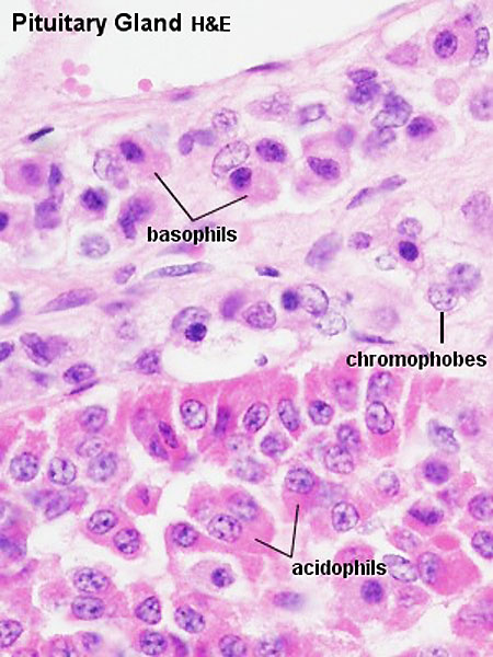 | 450 × 600 (72 KB) | Z8600021 (talk | contribs) | larger |
| 13:24, 5 October 2009 |  | 300 × 400 (45 KB) | S8600021 (talk | contribs) | Hya40he.jpg |
You cannot overwrite this file.