Category:Mouse E7.5: Difference between revisions
mNo edit summary |
mNo edit summary |
||
| (4 intermediate revisions by the same user not shown) | |||
| Line 7: | Line 7: | ||
{{Mouse}} | {{Mouse}} | ||
==Events== | ==Events== | ||
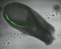
* {{gastrulation}} - p120-catenin regulates {{WNT}} signaling and EMT in the mouse embryo. "{{epithelial mesenchymal transition}}s (EMTs) require a complete reorganization of cadherin-based cell-cell junctions. p120-catenin binds to the cytoplasmic juxtamembrane domain of classical cadherins and regulates their stability, suggesting that p120-catenin may play an important role in EMTs. Here, we describe the role of p120-catenin in {{mouse}} {{gastrulation}}, an EMT that can be imaged at cellular resolution and is accessible to genetic manipulation. Mouse embryos that lack all p120-catenin, or that lack p120-catenin in the embryo proper, survive to midgestation. However, mutants have specific defects in gastrulation, including a high rate of p53-dependent cell death, a bifurcation of the posterior axis, and defects in the migration of mesoderm; all are associated with abnormalities in the primitive streak, the site of the EMT. In embryonic day 7.5 (E7.5) mutants, the domain of expression of the streak marker Brachyury (T) expands more than 3-fold, from a narrow strip of posterior cells to encompass more than one-quarter of the embryo. After {{ME7.5}}, the enlarged T+ domain splits in 2, separated by a mass of {{mesoderm}} cells. Brachyury is a direct target of canonical WNT signaling, and the domain of {{WNT}} response in p120-catenin mutant embryos, like the T domain, is first expanded, and then split, and high levels of nuclear β-catenin levels are present in the cells of the posterior embryo that are exposed to high levels of WNT ligand. The data suggest that p120-catenin stabilizes the membrane association of β-catenin, thereby preventing accumulation of nuclear β-catenin and excessive activation of the WNT pathway during EMT."{{#pmid:31371508|PMID31371508}} | |||
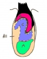
* {{Heart}} - bilateral paired cardiogenic plates (myocardial primordia).{{#pmid:19359541|PMID19359541}} | |||
===References=== | ===References=== | ||
<references/> | <references/> | ||
'''Search Pubmed:''' [http://www.ncbi.nlm.nih.gov/sites/entrez?db=pubmed&cmd=search&term=Mouse+E7.5 Mouse E7.5] | |||
Latest revision as of 08:28, 20 November 2019

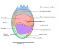
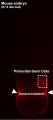
The Embryology pages and media listed below relate to mouse embryonic day 7.5 E7.5 of development. This staging by "days" relate to in the female presence of a vaginal plug indicating that the mating occurred, see timed pregnancy.
- Mouse Stages: E1 | E2.5 | E3.0 | E3.5 | E4.5 | E5.0 | E5.5 | E6.0 | E7.0 | E7.5 | E8.0 | E8.5 | E9.0 | E9.5 | E10 | E10.5 | E11 | E11.5 | E12 | E12.5 | E13 | E13.5 | E14 | E14.5 | E15 | E15.5 | E16 | E16.5 | E17 | E17.5 | E18 | E18.5 | E19 | E20 | Timeline | About timed pregnancy
| Carnegie | Stage | |||||||||||||||||||||||
| Human | Days | 1 | 2-3 | 4-5 | 5-6 | 7-12 | 13-15 | 15-17 | 17-19 | 20 | 22 | 24 | 28 | 30 | 33 | 36 | 40 | 42 | 44 | 48 | 52 | 54 | 55 | 58 |
| Mouse | Days | 1 | 2 | 3 | E4.5 | E5.0 | E6.0 | E7.0 | E8.0 | E9.0 | E9.5 | E10 | E10.5 | E11 | E11.5 | E12 | E12.5 | E13 | E13.5 | E14 | E14.5 | E15 | E15.5 | E16 |
| Rat | Days | 1 | 3.5 | 4-5 | 5 | 6 | 7.5 | 8.5 | 9 | 10.5 | 11 | 11.5 | 12 | 12.5 | 13 | 13.5 | 14 | 14.5 | 15 | 15.5 | 16 | 16.5 | 17 | 17.5 |
| Note these Carnegie stages are only approximate day timings for average of embryos. Links: Carnegie Stage Comparison | ||||||||||||||||||||||||
| ||||||||||||||||||||||||
| Timeline Links: human timeline | mouse timeline | mouse detailed timeline | chicken timeline | rat timeline | Medaka | Category:Timeline |
Events
- gastrulation - p120-catenin regulates WNT signaling and EMT in the mouse embryo. "epithelial mesenchymal transitions (EMTs) require a complete reorganization of cadherin-based cell-cell junctions. p120-catenin binds to the cytoplasmic juxtamembrane domain of classical cadherins and regulates their stability, suggesting that p120-catenin may play an important role in EMTs. Here, we describe the role of p120-catenin in mouse gastrulation, an EMT that can be imaged at cellular resolution and is accessible to genetic manipulation. Mouse embryos that lack all p120-catenin, or that lack p120-catenin in the embryo proper, survive to midgestation. However, mutants have specific defects in gastrulation, including a high rate of p53-dependent cell death, a bifurcation of the posterior axis, and defects in the migration of mesoderm; all are associated with abnormalities in the primitive streak, the site of the EMT. In embryonic day 7.5 (E7.5) mutants, the domain of expression of the streak marker Brachyury (T) expands more than 3-fold, from a narrow strip of posterior cells to encompass more than one-quarter of the embryo. After E7.5, the enlarged T+ domain splits in 2, separated by a mass of mesoderm cells. Brachyury is a direct target of canonical WNT signaling, and the domain of WNT response in p120-catenin mutant embryos, like the T domain, is first expanded, and then split, and high levels of nuclear β-catenin levels are present in the cells of the posterior embryo that are exposed to high levels of WNT ligand. The data suggest that p120-catenin stabilizes the membrane association of β-catenin, thereby preventing accumulation of nuclear β-catenin and excessive activation of the WNT pathway during EMT."[1]
References
- ↑ Hernández-Martínez R, Ramkumar N & Anderson KV. (2019). p120-catenin regulates WNT signaling and EMT in the mouse embryo. Proc. Natl. Acad. Sci. U.S.A. , 116, 16872-16881. PMID: 31371508 DOI.
- ↑ Savolainen SM, Foley JF & Elmore SA. (2009). Histology atlas of the developing mouse heart with emphasis on E11.5 to E18.5. Toxicol Pathol , 37, 395-414. PMID: 19359541 DOI.
Search Pubmed: Mouse E7.5
Media in category 'Mouse E7.5'
The following 11 files are in this category, out of 11 total.
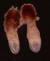
 Brachyury expression in 7.5dpc CD1 mouse embryos.jpg 386 × 313; 14 KB
 Day 7.5 Neural plate, presomite stage.JPG 425 × 537; 23 KB
 Mouse pax7 neural fold 01.jpg 513 × 915; 100 KB
 Mouse REN expression 01.jpg 904 × 1,000; 293 KB
 Mouse theiler stage 11.JPG 689 × 567; 42 KB
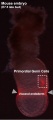
 Mouse- E7.5 early bud.jpg 568 × 698; 49 KB
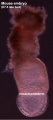
 Mouse- E7.5 early late.jpg 490 × 1,108; 68 KB
 Mouse- E7.5 late bud 01.jpg 442 × 1,000; 52 KB
 Mouse- E7.5 late bud 02.jpg 442 × 1,000; 77 KB
 Mouse- E7.5 late bud 03.jpg 442 × 1,000; 50 KB
 Mouse- E7.5 late bud.jpg 490 × 1,108; 68 KB


