Category:Carnegie Embryo 8963
From Embryology

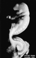
This Embryology category shows pages and images that relate to the Carnegie Collection Embryo No.8963. This 22 somite embryo was a tubal pregnancy originally classified as University of Chicago No. H1093. Described by Wen (1928)[1] with particular reference to the neural, renal, vascular systems.
Carnegie stage 12 - Week 4, 26 - 30 days, GA - week 6, 3.8 mm. (dors. flex.), Somite Number 22. 14.5 x 11 x 7 mm.
| Serial No. | Pairs of somites | Size (mm) | Grade | Fixative | Embedding Medium | Plane | Thinness (µm) | Stain | Year | Notes |
|---|---|---|---|---|---|---|---|---|---|---|
| 8963 | 22 | E, 3.8 Ch , 14.5 | Fair | Formol | C-P | Trans | 10 | I.H. | 1928 | Univ. Chicago. No. H 1093 Studied by Wen (1928)[1] |
| Carnegie Collection Embryos - Stage 12 | ||||||||||
|---|---|---|---|---|---|---|---|---|---|---|
| Serial No. | Pairs of somites | Size (mm) | Grade | Fixative | Embedding Medium | Plane | Thinness (µm) | Stain | Year | Notes |
| 209 | ca_24 | EH3 Ch.,15 | Poor | Alc | P | Coronal | 50 | Al. coch. | 1902 | |
| 250 | 19? | E , 2 (11, 10x9x9 | Poor | p | ? | Sagittal | 20 | Al. coch. | p | |
| 384 | P | E 2_5 Ch.,13 | Poor | Formalin | P | Transverse | 10 | H.&E. | 1907 | Macerated. Narrow yolk stalk |
| 486 | 21 | E.,4 Ch., 22 | Good | Corros. acetic | P | Transverse | 10 | Al. Coch. | 1911 | |
| 1062 | 29 | E.,4.5 Ch., 20 | Good | Formalin | P | Transverse | 20 | Al. coch. | 1915 | Transitional to next stage |
| 2197 | ? | E_,5_3 Ch., 19.5 | Poor | Formalin | P | Transverse | 10 | Al. coch, or. G. | 1918 | |
| 4245-7 | ca.24 | E.,3.5 Ch., 24 | Good | Alc, formol | P | Transverse | 10 | Al. coch. | 1923 | Caudal neuropore widely open |
| 4479 | P | E.,5 .8 Ch , 17 | Poor | Formalin | P | Transverse | 40 | Al. coch. | 1923 | Macerated. Upper lirnb buds not visible |
| 4736 | 26 | E.,3.0 Ch.,20 | Good | Formalin | P | Coronal | 10 | Al. coch. | 1924 | No upper limb buds. Caudal neuropore closed |
| 4759 | ? | E.,4.5 Ch.,15 | Good | Formalin | P | Transverse | 15 | (Stain - Haematoxylin Eosin) | 1924 | Neural tube folded |
| 4784 | 23 | E_,3 | Good | P | P | Transverse | 10 | p | 1924 | |
| 5035 | 25-28 | E.,3.8 Ch.,18 | Good | Formalin | C-P | Transverse | 10 | Al. coch. | 1925 | |
| 5048 | ca_25 | E_,3_5 | Good | Formalin | C-P | Transverse | 10 | Al. coch. | 1925 | Tubal Injured |
| 5056 | 25 | E,,3 Ch.,12 | Good | Formalin | P | Transverse | 10 | Al. coch. | 1925 | |
| 5206 | ? | E.,4 Ch., 51x31x30 | Poor | ?? | P | Transverse | 20 | Al. coch. | 1926 | Tubal |
| 5300 | ? | E., 4.5 Ch,16.5 | Poor | Formalin | P | Transverse | 20 | Al. coch. | 1926 | Autopsy. Partly macerated |
| 5923 | 28 | E.,4 Ch.,15 | Exc. | Formalin | P | Transverse | 10 | Al. coch. | 1929 | |
| 6097 | 25 | E,3.4 Ch., 12.5 | Exc. | Formalin | C-P | Transverse | 10 | Al. coch, eosin | 1930 | Tubal Ag added to slides 1-3 |
| 6144 | 27 | E. 3.3 Ch.,11 | Good | Lysol—Zenker | C—P | Transverse | 10 | Al. coch. | 1930 | |
| 6488 | 28 | E, 32 Ch,22 | Good | Formalin | C—P | Transverse | 10 | Al. coch. | 1932 | |
| 6937 | 26 | E.,3 Ch , 12 | Poor | Formalin | C—P | Coronal | 10 | I.H.,or.G. | 1934 | Tubal Caudal neuropore closed |
| 7724 | ca.29 | E,3.5 Ch.,18 | Good | Formalin | C—P | Sagittal | 8 | (Stain - Haematoxylin Eosin) | 1940 | Caudal end broken |
| 7852 | 25 | E , 3.7 Ch,26 | Exc. | Formalin | C—P | Transverse | 10 | (Stain - Haematoxylin Eosin) | 1940 | Typical for stage 12 |
| 7999 | ca.28 | E,3.2 Ch , 15 | Exc. | Bouin | C-P | Transverse | 10 | (Stain - Haematoxylin Eosin) | 1942 | Caudal defect |
| 8505a | 24 | Ch, 23.5 | Exc. | Formalin | C-P | Transverse | P | H. Phlox. | 1947 | |
| 8505b | 23 | Ch,24 | Exc. | Formalin | C-P | Sagittal | p | Azan | 1947 | Twins |
| 8941 | 28 | E,4.9 Ch, 35 | Exc. | Zenker | C-P | Transverse | 6 I.H. | 1927 | Univ. Chicago No. H 1261 | |
| 8942 | 25 | E, 38 Ch, 35 | Exc. | Formal-Zenker | C-P | Coronal | 5 11-1. | 1930 | Univ. Chicago No. H 1382 | |
| 8943 | 22 | E. 3.9 Ch, 20.4 | Exc. | Formal-Zenker | C-P | Transverse | 8 | H.&E. | 1934 | Univ. Chicago No. H 1481 |
| 8944 | 25 | E,4 Ch,,25 | Exc. | Formal-Zenker | C-P | Sagittal | 8 | I.H. | 1936 | Univ. Chicago No. H 1514 |
| 8963 | 22 | E, 3.8 Ch , 14.5 | Fair | Formalin | C-P | Transverse | 10 | I.H. | 1928 | Univ. Chicago. No. H 1093 Studied by Wen (1928)[1] |
| 8964 | 23 | E,2.8 Ch - 25 | Poor | Formalin | p | Transverse | 8 | I.H. | 1928 | Univ_ Chicago No. H984 Studied by Wen (1928)[1] |
| 9154 | 24 | E, 5 4 | Exc | Formalin | C-P | Transverse | I.H. & phlox. | 1953 | ||
Abbreviations
| ||||||||||
References
| ||||||||||
The embryo was sectioned into 323 sections at 10 µ.
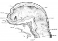
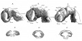
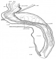
- Embryo H1093 (fig. 1, B) was obtained by Dr. Garrette Van Sweringen and Dr. W. W. Duemling, of Fort Wayne, Indiana. The clinical record gives the following points: “The October period began on the 6th, 1924; next period from 10th to 14th or 15th. The expected period on December 10th did not appear. Flow supposed to be menstruation appeared on December 22nd, but bloody discharge continued until 31st. Contraceptive measures were employed until after November period. Symptoms of ectopic pregnancy were observed on January 4, 1925, and operation was performed on the same day.” In cutting open the hematoma, the head of the embryo was severed from the trunk just caudal to the otocyst. Although no loss of tissue is apparently involved, yet the difference in the plane of sections of the two portions of the embryo makes it materially difficult to follow the series in the region of rhombomere VI. The specimen was fixed in neutral formalin, double embedded, cut transversely, and stained with iron hematoxylin. Histological differentiation is fair, but there is much shrinkage. Cells with coarse granules evidently representing necrobiosis are found abundant in the neural crest as well as the medullary tube. A wax model of the nervous system in front of the otocyst was made from serial photomicrographs at the magnification of 150 times.[1]
- ↑ Wen, I. C., The anatomy of human embryos with seventeen to twenty-three pairs of somites J. Comp. Neural, 1928, 45:301-376.
Pages in category 'Carnegie Embryo 8963'
This category contains only the following page.
Media in category 'Carnegie Embryo 8963'
The following 6 files are in this category, out of 6 total.
 Wen1928-Fig01b.jpg 471 × 759; 31 KB
 Wen1928-Fig06.jpg 1,280 × 917; 280 KB
 Wen1928-Fig07.jpg 1,265 × 648; 166 KB
 Wen1928-Fig19.jpg 1,200 × 1,259; 238 KB
 Wen1928-Fig26.jpg 850 × 800; 173 KB
 Wen1928-Fig29.jpg 1,200 × 884; 133 KB


