File:Pituitary histology 001.jpg
Pituitary_histology_001.jpg (450 × 600 pixels, file size: 72 KB, MIME type: image/jpeg)
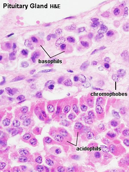
Pituitary Histology - Adenohypophysis
Pituitary, sheep - H&E
Acidophils are dark pink and basophils look light pink/blue.
- Pituitary Histology: Pituitary overview | Anterior H&E | Anterior H&E | Anterior labeled | PAS/O Overview | Acidophils | Basophils | Posterior labeled | Posterior unlabeled | Histology Stains | BGD - Endocrine Histology | Pituitary Development
Links: Histology | Histology Stains | Blue Histology images copyright Lutz Slomianka 1998-2009. The literary and artistic works on the original Blue Histology website may be reproduced, adapted, published and distributed for non-commercial purposes. See also the page Histology Stains.
Cite this page: Hill, M.A. (2024, June 1) Embryology Pituitary histology 001.jpg. Retrieved from https://embryology.med.unsw.edu.au/embryology/index.php/File:Pituitary_histology_001.jpg
- © Dr Mark Hill 2024, UNSW Embryology ISBN: 978 0 7334 2609 4 - UNSW CRICOS Provider Code No. 00098G
Original File Name: Hya40he.jpg
File history
Click on a date/time to view the file as it appeared at that time.
| Date/Time | Thumbnail | Dimensions | User | Comment | |
|---|---|---|---|---|---|
| current | 14:29, 12 May 2012 | | 450 × 600 (72 KB) | Z8600021 (talk | contribs) | larger |
| 13:24, 5 October 2009 |  | 300 × 400 (45 KB) | S8600021 (talk | contribs) | Hya40he.jpg |
You cannot overwrite this file.